1MUQ image
Deposition Date 2002-09-24
Release Date 2003-07-01
Last Version Date 2024-11-20
Entry Detail
PDB ID:
1MUQ
Title:
X-ray Crystal Structure of Rattlesnake Venom Complexed With Thiodigalactoside
Biological Source:
Source Organism(s):
Crotalus atrox (Taxon ID: 8730)
Method Details:
Experimental Method:
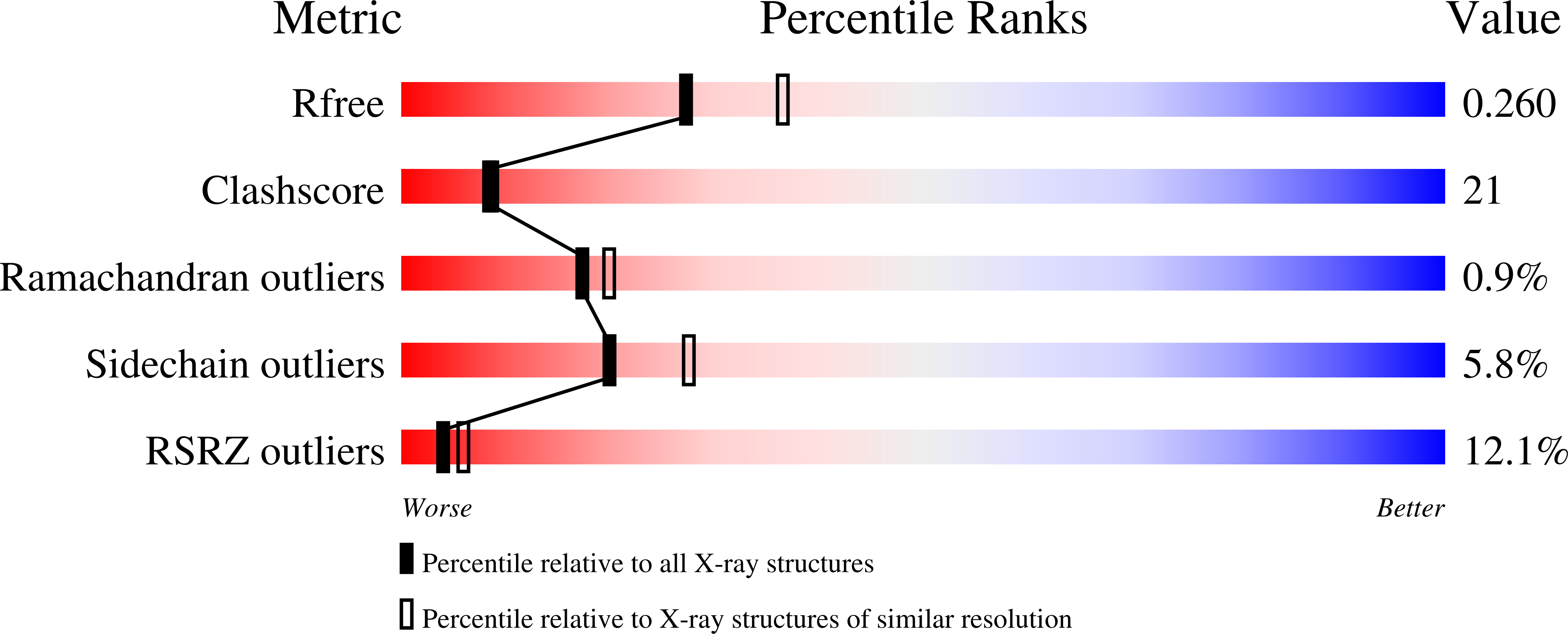
Resolution:
2.30 Å
R-Value Free:
0.25
R-Value Work:
0.21
R-Value Observed:
0.21
Space Group:
C 2 2 21
Macromolecular Entities
Structures with similar UniProt ID
Protein Blast
Polymer Type:polypeptide(L)
Molecule:Galactose-specific lectin
Chain IDs:A, B, C, D, E
Chain Length:135
Number of Molecules:5
Biological Source:Crotalus atrox
Peptide-like Molecules
PRD_900027
Primary Citation
X-ray Crystal Structure of a Galactose-Specific C-Type Lectin Possessing a Novel Decameric Quaternary Structure.
Biochemistry 43 3783 3792 (2004)
PMID: 15049685 DOI: 10.1021/bi035871a

Abstact

Rattlesnake venom lectin (RSL) from the western diamondback rattlesnake (Crotalus atrox) is an oligomeric galactose-specific C-type lectin. The X-ray crystal structure of RSL, in complex with lactose and thiodigalactoside, at 2.2 and 2.3 A resolution, respectively, reveals a decameric protein composed of two 5-fold symmetric pentamers arranged in a staggered, back-to-back orientation. Each monomer corresponds to a single canonical C-type lectin carbohydrate recognition domain devoid of accessory domains and is disulfide-bonded to a monomer in the other pentamer. The structure is the first example of that of a carbohydrate complex of a vertebrate galactose-specific C-type lectin. The 10 carbohydrate-binding sites, located on the rim of the decamer, suggest a role for multivalent interactions and a mechanism for RSL's ability to promote receptor cross-linking and cell aggregation.

Legend

Protein

Chemical

Disease

Primary Citation of related structures
Feedback Form
Name
Email
Institute
Feedback