1MUM image
Deposition Date 2002-09-24
Release Date 2003-04-29
Last Version Date 2024-03-13
Entry Detail
PDB ID:
1MUM
Keywords:
Title:
Structure of the 2-Methylisocitrate Lyase (PrpB) from Escherichia coli
Biological Source:
Source Organism:
Escherichia coli (Taxon ID: 562)
Host Organism:
Method Details:
Experimental Method:
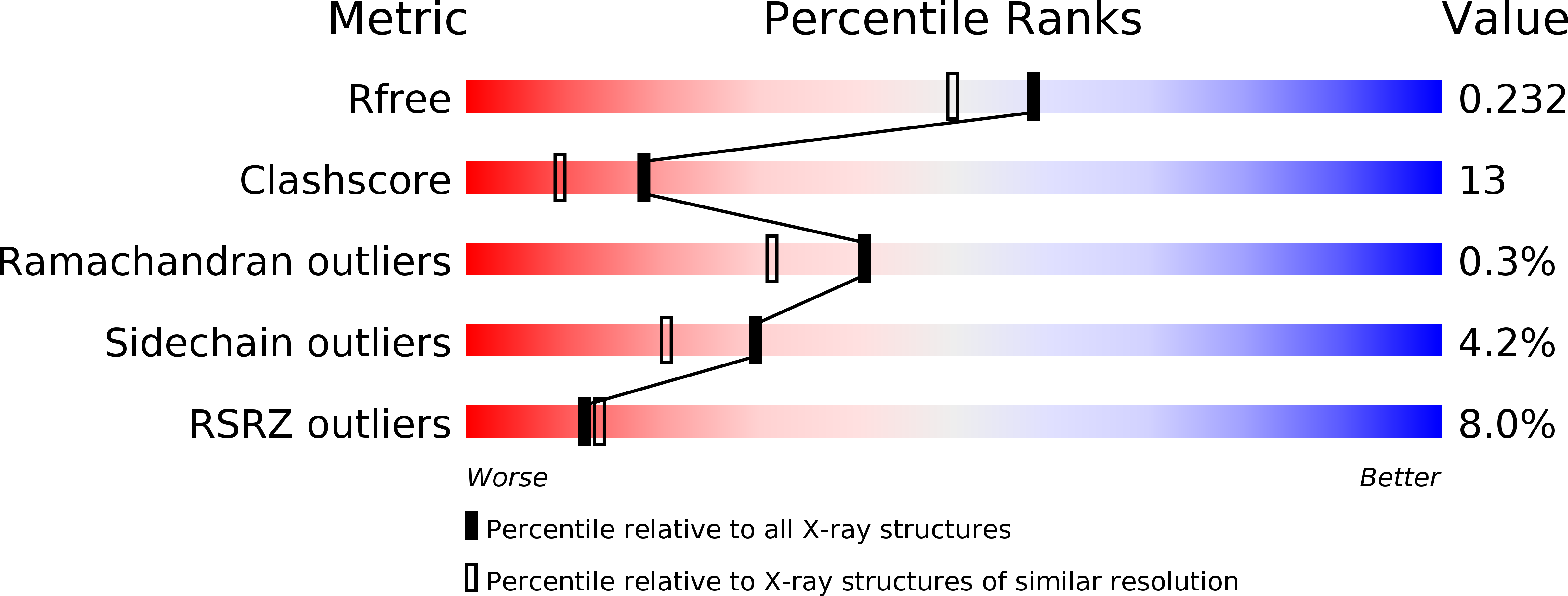
Resolution:
1.90 Å
R-Value Free:
0.23
R-Value Work:
0.21
R-Value Observed:
0.21
Space Group:
P 32 2 1
Macromolecular Entities
Structures with similar UniProt ID
Protein Blast
Polymer Type:polypeptide(L)
Molecule:2-methylisocitrate lyase
Gene (Uniprot):prpB
Chain IDs:A, B
Chain Length:295
Number of Molecules:2
Biological Source:Escherichia coli
Ligand Molecules
Primary Citation
Crystal structure of 2-methylisocitrate lyase (PrpB) from Escherichia coli and modelling of its ligand bound active centre.
J.Mol.Biol. 328 609 621 (2003)
PMID: 12706720 DOI: 10.1016/S0022-2836(03)00358-9

Abstact

Following acetate, propionate is the second most abundant low molecular mass carbon compound found in soil. Many microorganisms, including most, if not all fungi, as well as several aerobic bacteria, such as Escherichia coli and Salmonella enterica oxidize propionate via the methylcitrate cycle. The enzyme 2-methylisocitrate lyase (PrpB) from Escherichia coli catalysing the last step of this cycle, the cleavage of 2-methylisocitrate to pyruvate and succinate, was crystallised and its structure determined to a resolution of 1.9A. The enzyme, which strictly depends on Mg(2+) for catalysis, belongs to the isocitrate lyase protein family. A common feature of members of this enzyme family is the movement of a so-called "active site loop" from an open into a closed conformation upon substrate binding thus shielding the reactants from the surrounding solvent. Since in the presented structure, PrpB contains, apart from a Mg(2+), no ligand, the active site loop is found in an open conformation. This conformation, however, differs significantly from the open conformation present in the so far known structures of ligand-free isocitrate lyases. A possible impact of this observation with respect to the different responses of isocitrate lyases and PrpB upon treatment with the common inhibitor 3-bromopyruvate is discussed. Based on the structure of ligand-bound isocitrate lyase from Mycobacterium tuberculosis a model of the substrate-bound PrpB enzyme in its closed conformation was created which provides hints towards the substrate specificity of this enzyme.

Legend

Protein

Chemical

Disease

Primary Citation of related structures