1MUI image
Deposition Date 2002-09-23
Release Date 2002-10-23
Last Version Date 2024-02-14
Entry Detail
PDB ID:
1MUI
Keywords:
Title:
Crystal structure of HIV-1 protease complexed with Lopinavir.
Biological Source:
Source Organism:
Host Organism:
Method Details:
Experimental Method:
Resolution:
2.80 Å
R-Value Free:
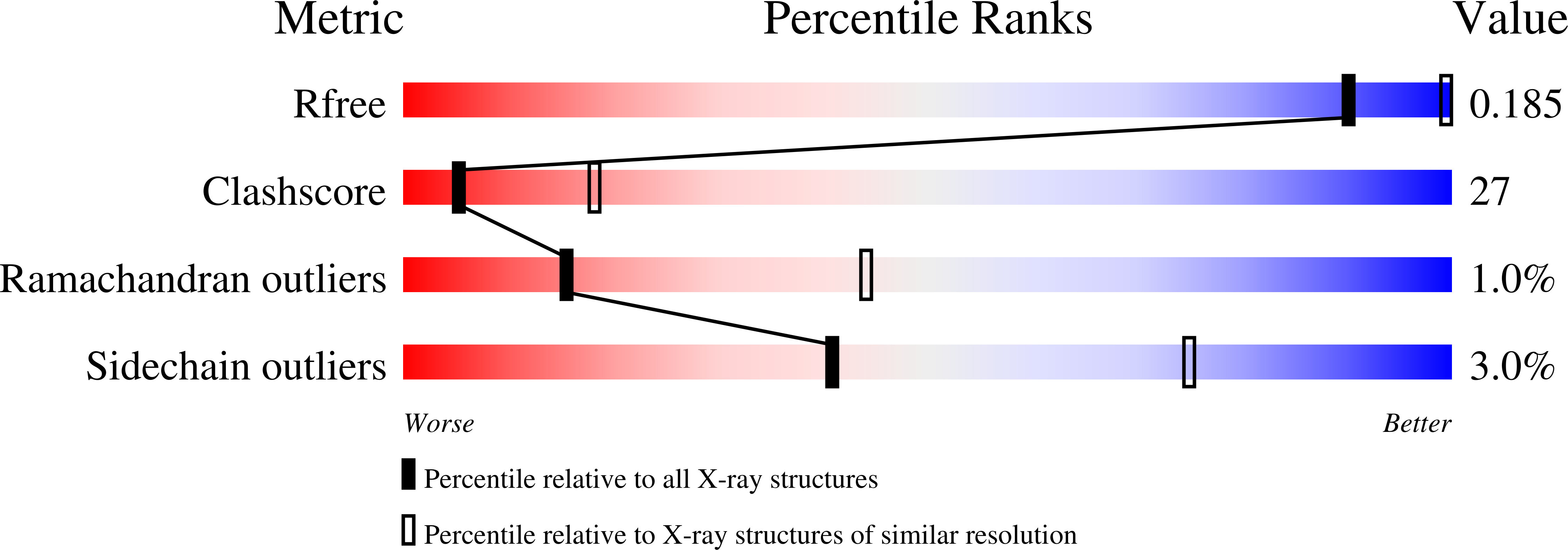
0.32
R-Value Work:
0.26
R-Value Observed:
0.30
Space Group:
P 61
Macromolecular Entities
Polymer Type:polypeptide(L)
Molecule:protease
Mutations:N37S
Chain IDs:A (auth: B), B (auth: A)
Chain Length:99
Number of Molecules:2
Biological Source:Human immunodeficiency virus 1
Ligand Molecules
Primary Citation
X-ray Crystallographic Structure of ABT-378 (Lopinavir) Bound to HIV-1 Protease
BIOORG.MED.CHEM. 10 2803 2806 (2002)
PMID: 12057670 DOI: 10.1016/S0968-0896(02)00051-2

Abstact

The crystal structure of ABT-378 (lopinavir), bound to the active site of HIV-1 protease is described. A comparison with crystal structures of ritonavir, A-78791, and BILA-2450 shows some analogous features with previous reported compounds. A cyclic urea unit in the P(2) position of ABT-378 is novel and makes two bidentate hydrogen bonds with Asp 29 of HIV-1 protease. In addition, a previously unreported shift in the Gly 48 carbonyl position is observed. A discussion of the structural features responsible for its high potency against wild-type HIV protease is given along with an analysis of the effect of active site mutations on potency in in vitro assays.

Legend

Protein

Chemical

Disease

Primary Citation of related structures