1MU7 image
Deposition Date 2002-09-23
Release Date 2003-01-07
Last Version Date 2024-02-14
Entry Detail
PDB ID:
1MU7
Keywords:
Title:
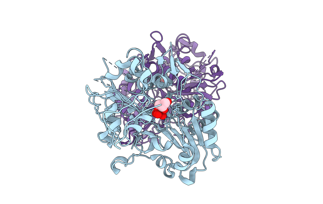
Crystal Structure of a Human Tyrosyl-DNA Phosphodiesterase (Tdp1)-Tungstate Complex
Biological Source:
Source Organism:
Homo sapiens (Taxon ID: 9606)
Host Organism:
Method Details:
Experimental Method:
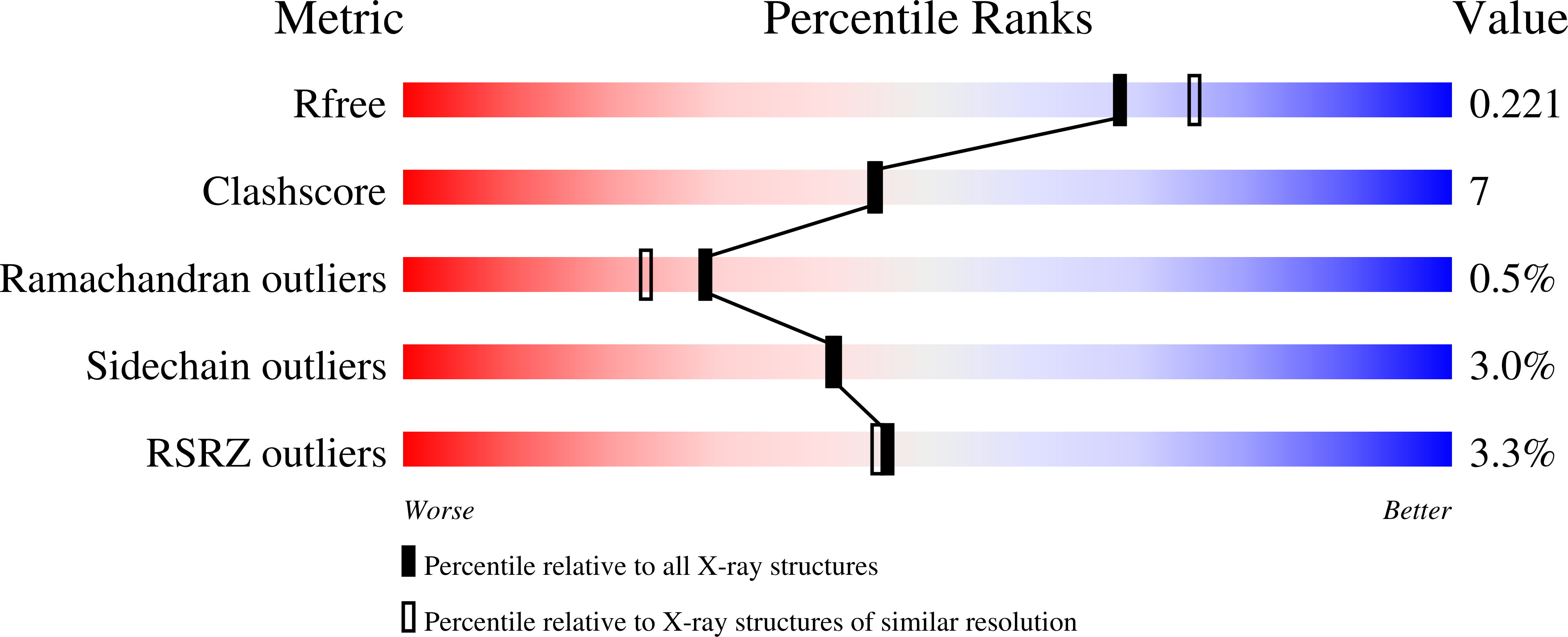
Resolution:
2.00 Å
R-Value Free:
0.21
R-Value Work:
0.20
R-Value Observed:
0.20
Space Group:
P 21 21 21
Macromolecular Entities
Structures with similar UniProt ID
Protein Blast
Polymer Type:polypeptide(L)
Molecule:Tyrosyl-DNA Phosphodiesterase
Mutagens:D322N,M328T,F548L
Chain IDs:A, B
Chain Length:485
Number of Molecules:2
Biological Source:Homo sapiens
Primary Citation
Insights Into Substrate Binding and Catalytic Mechanism of Human Tyrosyl-DNA Phosphodiesterase (Tdp1) from Vanadate- and Tungstate-Inhibited Structures
J.Mol.Biol. 324 917 932 (2003)
PMID: 12470949 DOI: 10.1016/S0022-2836(02)01154-3

Abstact

Tyrosyl-DNA phosphodiesterase (Tdp1) is a DNA repair enzyme that catalyzes the hydrolysis of a phosphodiester bond between a tyrosine residue and a DNA 3'-phosphate. The only known example of such a linkage in eukaryotic cells occurs normally as a transient link between a type IB topoisomerase and DNA. Thus human Tdp1 is thought to be responsible for repairing lesions that occur when topoisomerase I becomes stalled on the DNA in the cell. Tdp1 has also been shown to remove glycolate from single-stranded DNA containing a 3'-phosphoglycolate, suggesting a role for Tdp1 in repair of free-radical mediated DNA double-strand breaks. We report the three-dimensional structures of human Tdp1 bound to the phosphate transition state analogs vanadate and tungstate. Each structure shows the inhibitor covalently bound to His263, confirming that this residue is the nucleophile in the first step of the catalytic reaction. Vanadate in the Tdp1-vanadate structure has a trigonal bipyramidal geometry that mimics the transition state for hydrolysis of a phosphodiester bond, while Tdp1-tungstate displays unusual octahedral coordination. The presence of low-occupancy tungstate molecules along the narrow groove of the substrate binding cleft is suggestive evidence that this groove binds ssDNA. In both cases, glycerol from the cryoprotectant solution became liganded to the vanadate or tungstate inhibitor molecules in a bidentate 1,2-diol fashion. These structural models allow predictions to be made regarding the specific binding mode of the substrate and the mechanism of catalysis.

Legend

Protein

Chemical

Disease

Primary Citation of related structures
Feedback Form
Name
Email
Institute
Feedback