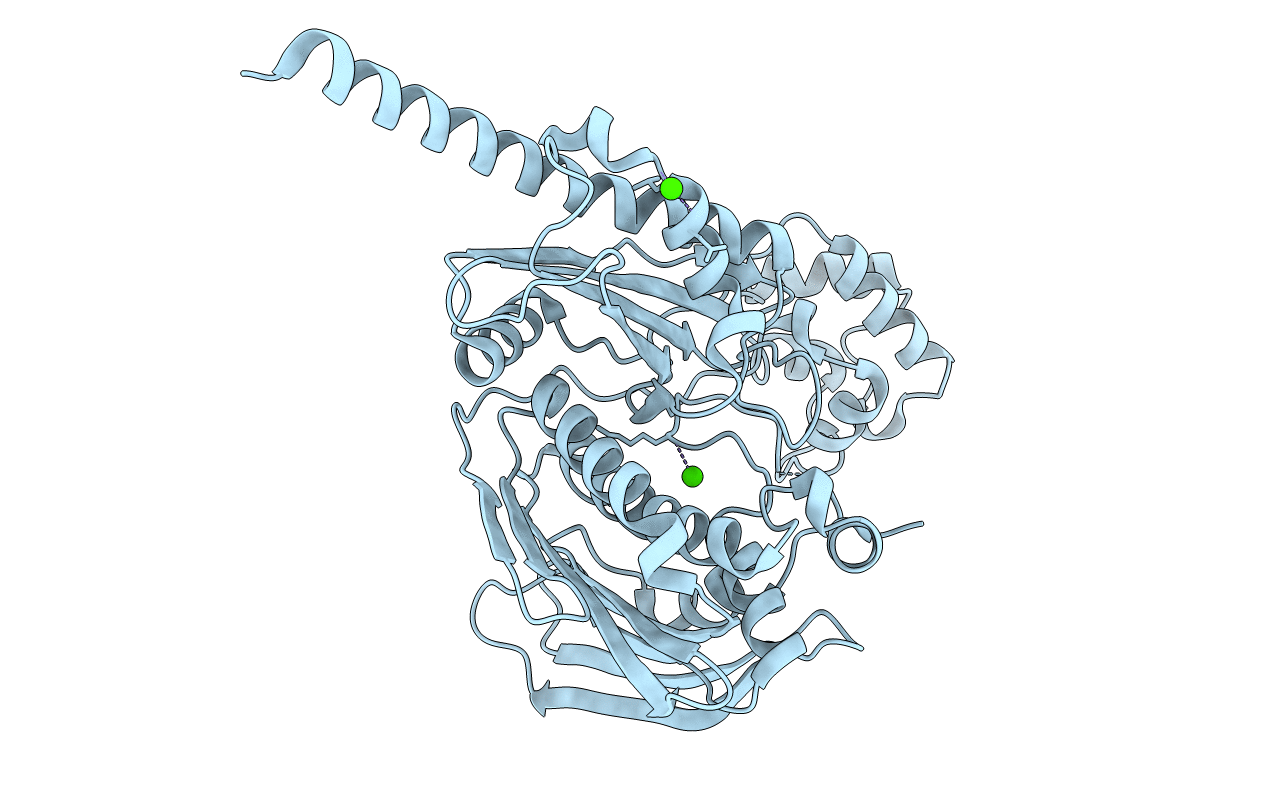
1MU5 image
Deposition Date 2002-09-23
Release Date 2003-01-07
Last Version Date 2024-02-14
Entry Detail
PDB ID:
1MU5
Keywords:
Title:
Structure of topoisomerase subunit
Biological Source:
Source Organism:
Host Organism:
Method Details:
Experimental Method:
Resolution:
2.00 Å
R-Value Free:
0.23
R-Value Work:
0.21
R-Value Observed:
0.21
Space Group:
P 21 21 2
Macromolecular Entities
Structures with similar UniProt ID
Protein Blast
Polymer Type:polypeptide(L)
Molecule:Type II DNA topoisomerase VI Subunit B
Gene (Uniprot):top6B
Chain IDs:A
Chain Length:471
Number of Molecules:1
Biological Source:Sulfolobus shibatae
Ligand Molecules
Primary Citation
Structure of the topoisomerase VI-B subunit: implications for type II topoisomerase mechanism and evolution
Embo J. 22 151 163 (2003)
PMID: 12505993 DOI: 10.1093/emboj/cdg008

Abstact

Type IIA and type IIB topoisomerases each possess the ability to pass one DNA duplex through another in an ATP-dependent manner. The role of ATP in the strand passage reaction is poorly understood, particularly for the type IIB (topoisomerase VI) family. We have solved the structure of the ATP-binding subunit of topoisomerase VI (topoVI-B) in two states: an unliganded monomer and a nucleotide-bound dimer. We find that topoVI-B is highly structurally homologous to the entire 40-43 kDa ATPase region of type IIA topoisomerases and MutL proteins. Nucleotide binding to topoVI-B leads to dimerization of the protein and causes dramatic conformational changes within each protomer. Our data demonstrate that type IIA and type IIB topoisomerases have descended from a common ancestor and reveal how ATP turnover generates structural signals in the reactions of both type II topoisomerase families. When combined with the structure of the A subunit to create a picture of the intact topoisomerase VI holoenzyme, the ATP-driven motions of topoVI-B reveal a simple mechanism for strand passage by the type IIB topoisomerases.

Legend

Protein

Chemical

Disease

Primary Citation of related structures