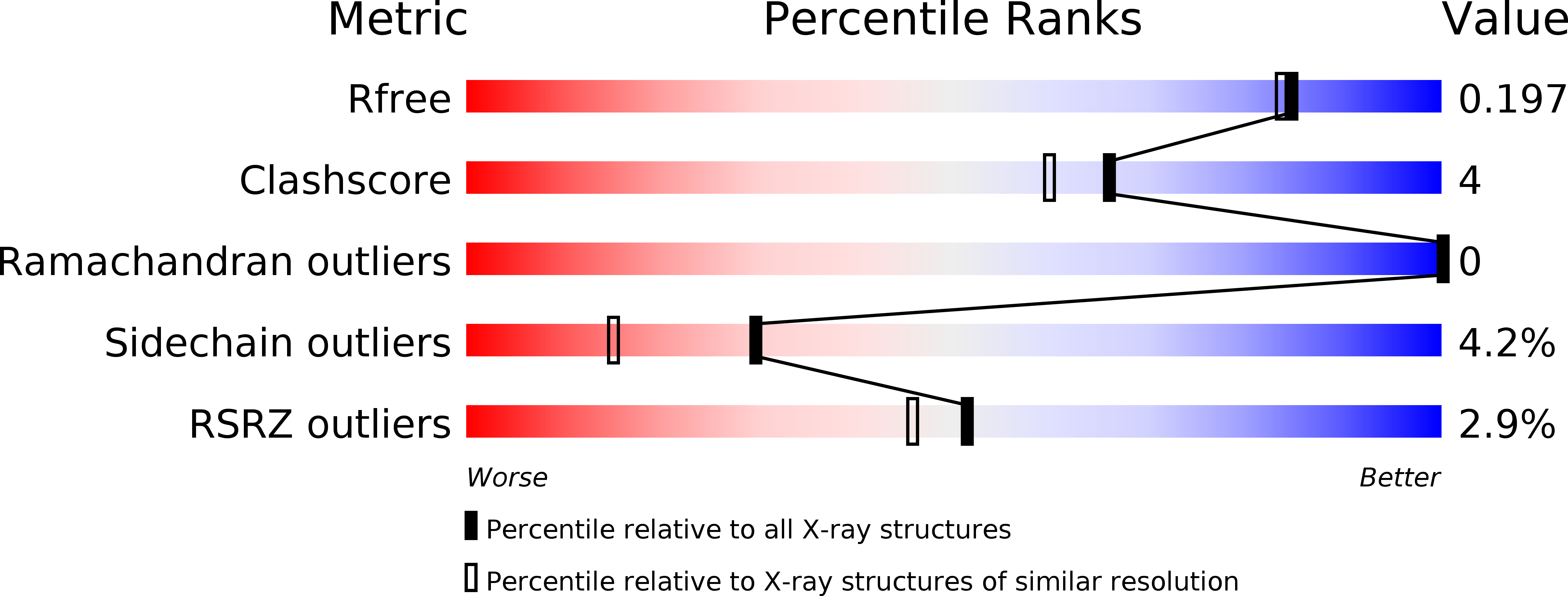
1MU4 image
Deposition Date 2002-09-23
Release Date 2003-10-07
Last Version Date 2024-02-14
Entry Detail
PDB ID:
1MU4
Title:
CRYSTAL STRUCTURE AT 1.8 ANGSTROMS OF THE BACILLUS SUBTILIS CATABOLITE REPRESSION HISTIDINE CONTAINING PROTEIN (CRH)
Biological Source:
Source Organism:
Host Organism:
Method Details:
Experimental Method:
Resolution:
1.80 Å
R-Value Free:
0.19
R-Value Work:
0.17
R-Value Observed:
0.17
Space Group:
P 43 2 2
Macromolecular Entities
Polymer Type:polypeptide(L)
Molecule:HPr-like protein crh
Gene (Uniprot):crh
Chain IDs:A, B
Chain Length:87
Number of Molecules:2
Biological Source:Bacillus subtilis
Ligand Molecules
Primary Citation
Dimerization of Crh by reversible 3D Domain Swapping Induces Structural Adjustments to its monomeric homologue HPR
J.Mol.Biol. 332 767 776 (2003)
PMID: 12972249 DOI: 10.1016/S0022-2836(03)00918-5

Abstact

The crystal structure of the regulatory protein Crh from Bacillus subtilis was solved at 1.8A resolution and showed an intertwined dimer formed by N-terminal beta1-strand swapping of two monomers. Comparison with the monomeric NMR structure of Crh revealed a domain swap induced conformational rearrangement of the putative interaction site with the repressor CcpA. The resulting conformation closely resembles that observed for the monomeric Crh homologue HPr, indicating that the Crh dimer is the active form binding to CcpA. An analogous dimer of HPr can be constructed without domain swapping, suggesting that HPr may dimerize upon binding to CcpA. Our data suggest that reversible 3D domain swapping of Crh might be an efficient regulatory mechanism to modulate its activity.

Legend

Protein

Chemical

Disease

Primary Citation of related structures