Deposition Date
2002-09-18
Release Date
2004-05-18
Last Version Date
2024-11-13
Entry Detail
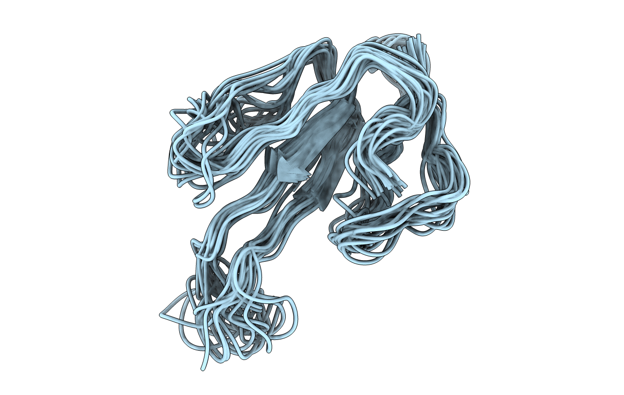
PDB ID:
1MR6
Keywords:
Title:
Solution Structure of gamma-Bungarotoxin:Implication for the role of the Residues Adjacent to RGD in Integrin Binding
Biological Source:
Source Organism(s):
Bungarus multicinctus (Taxon ID: 8616)
Method Details:
Experimental Method:
Conformers Calculated:
50
Conformers Submitted:
20
Selection Criteria:
structures with the least restraint violations, structures with the lowest energy