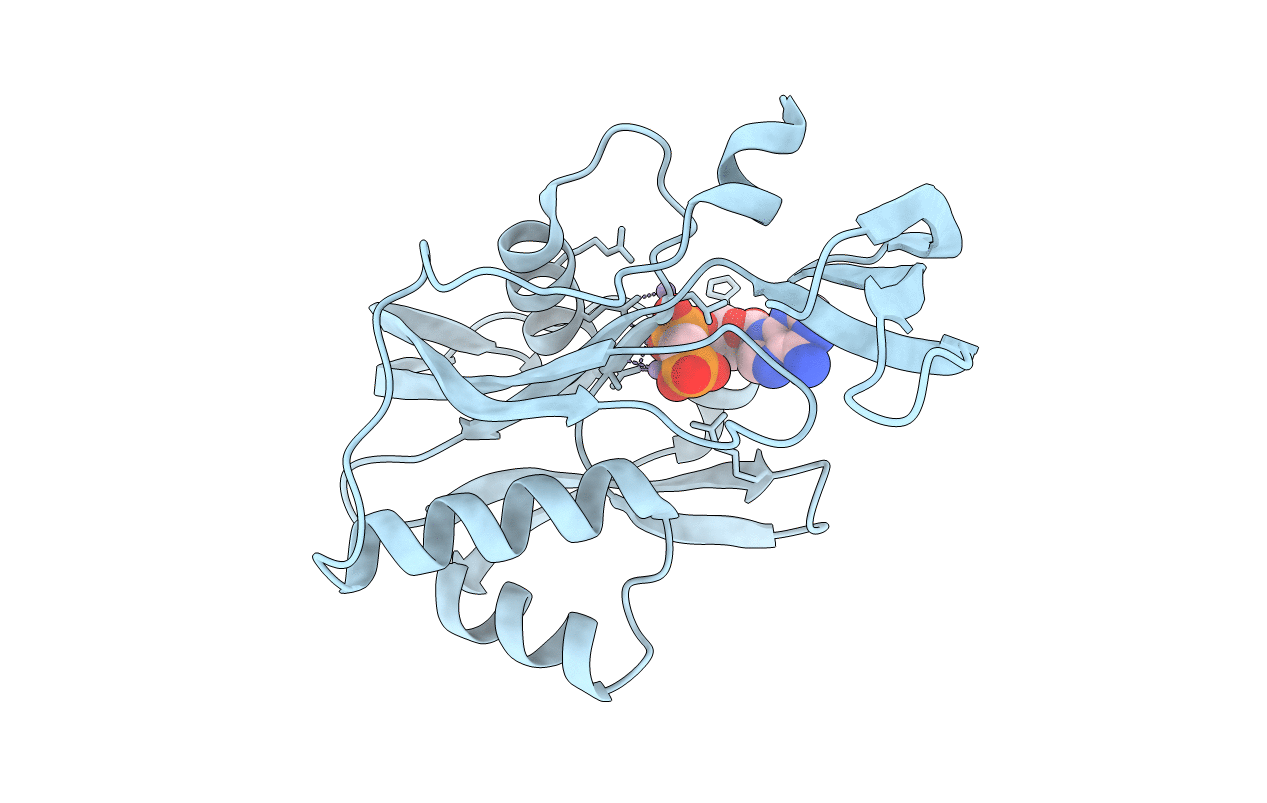
1MQW image
Deposition Date 2002-09-17
Release Date 2003-08-05
Last Version Date 2024-02-14
Entry Detail
PDB ID:
1MQW
Keywords:
Title:
Structure of the MT-ADPRase in complex with three Mn2+ ions and AMPCPR, a Nudix enzyme
Biological Source:
Source Organism:
Host Organism:
Method Details:
Experimental Method:
Resolution:
2.30 Å
R-Value Free:
0.26
R-Value Work:
0.20
Space Group:
P 61 2 2
Macromolecular Entities
Polymer Type:polypeptide(L)
Molecule:ADPR pyrophosphatase
Gene (Uniprot):MT1739
Chain IDs:A
Chain Length:207
Number of Molecules:1
Biological Source:Mycobacterium tuberculosis
Primary Citation
Structure and mechanism of MT-ADPRase, a Nudix hydrolase from Mycobacterium tuberculosis
Structure 11 1015 1023 (2003)
PMID: 12906832 DOI: 10.1016/S0969-2126(03)00154-0

Abstact

Nudix hydrolases are a family of proteins that contain the characteristic sequence GX(5)EX(7)REUXEEXG(I/L/V), the Nudix box. They catalyze the hydrolysis of a variety of nucleoside diphosphate derivatives such as ADP-ribose, Ap(n)A (3 </= n </= 6), NADH, and dATP. A number of Nudix hydrolases from several species, ranging from bacteria to humans, have been characterized, including, in some cases, the determination of their three-dimensional structures. The product of the Rv1700 gene of M. tuberculosis is a Nudix hydrolase specific for ADP-ribose (ADPR). We have determined the crystal structures of MT-ADPRase alone, and in complex with substrate, with substrate and the nonactivating metal ion Gd(3+), and in complex with a nonhydrolyzable ADPR analog and the activating metal ion Mn(2+). These structures, refined with data extending to resolutions between 2.0 and 2.3 A, showed that there are sequence differences in binding site residues between MT-ADPRase and a human homolog that may be exploited for antituberculosis drug development.

Legend

Protein

Chemical

Disease

Primary Citation of related structures