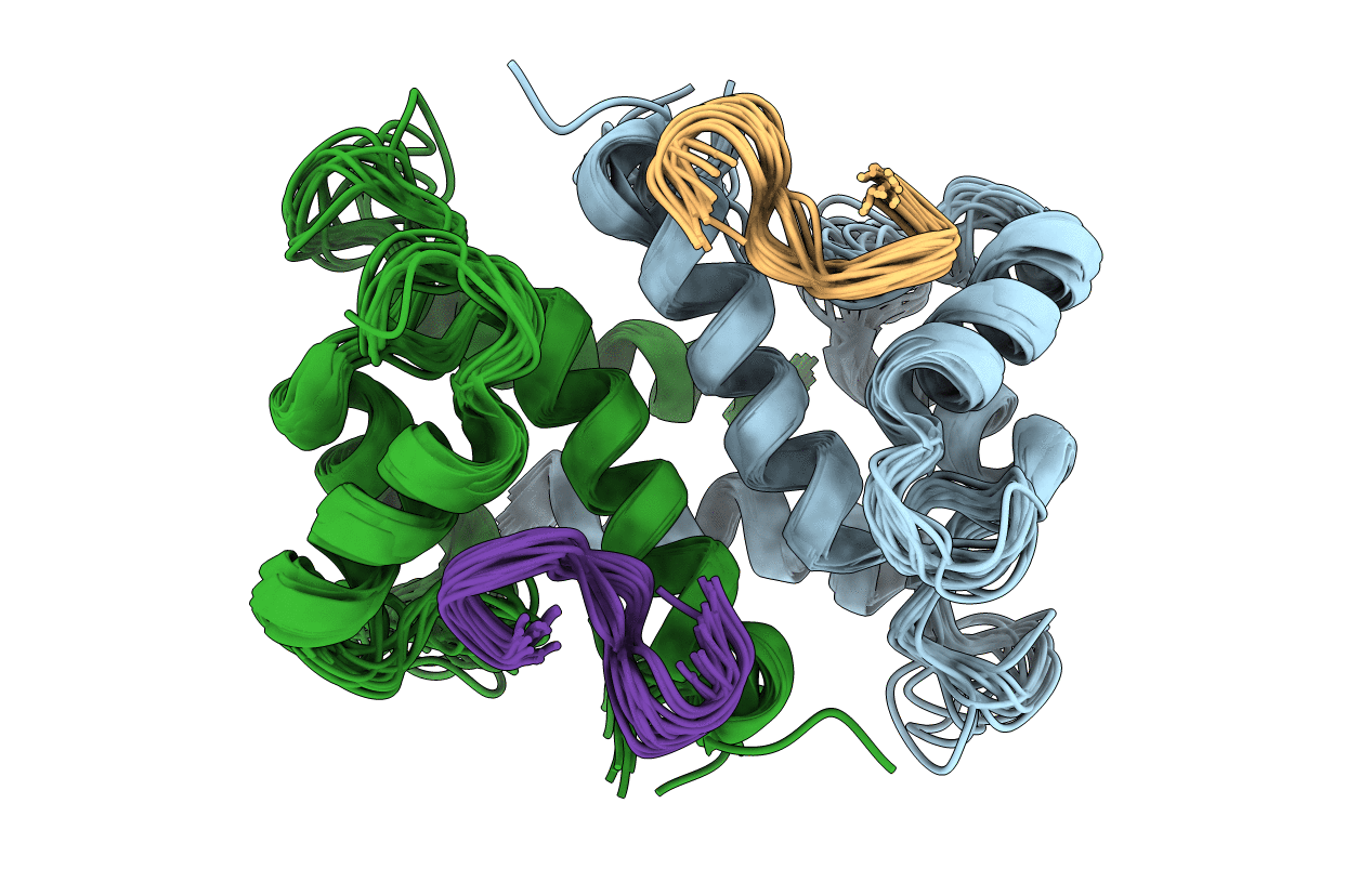
1MQ1 image
Deposition Date 2002-09-13
Release Date 2002-12-25
Last Version Date 2024-05-22
Entry Detail
PDB ID:
1MQ1
Title:
Ca2+-S100B-TRTK-12 complex
Biological Source:
Source Organism:
Homo sapiens (Taxon ID: 9606)
Host Organism:
Method Details:
Experimental Method:
Conformers Calculated:
80
Conformers Submitted:
17
Selection Criteria:
structures with the lowest energy
Macromolecular Entities
Structures with similar UniProt ID
Protein Blast
Polymer Type:polypeptide(L)
Molecule:S-100 protein, beta chain
Gene (Uniprot):S100B
Chain IDs:A, B
Chain Length:91
Number of Molecules:2
Biological Source:Homo sapiens
Structures with similar UniProt ID
Protein Blast
Polymer Type:polypeptide(L)
Molecule:F-actin capping protein alpha-1 subunit
Gene (Uniprot):CAPZA1
Chain IDs:C, D
Chain Length:12
Number of Molecules:2
Biological Source:
Ligand Molecules
Primary Citation
A novel S100 target conformation is revealed by the solution structure of the Ca2+-S100B-TRTK-12 complex.
J.Biol.Chem. 278 6251 6257 (2003)
PMID: 12480931 DOI: 10.1074/jbc.M210622200

Abstact

The Alzheimer-linked neural protein S100B is a signaling molecule shown to control the assembly of intermediate filament proteins in a calcium-sensitive manner. Upon binding calcium, a conformational change occurs in S100B exposing a hydrophobic surface for target protein interactions. The synthetic peptide TRTK-12 (TRTKIDWNKILS), derived from random bacteriophage library screening, bears sequence similarity to several intermediate filament proteins and has the highest calcium-dependent affinity of any target molecule for S100B to date (K(d) <1 microm). In this work, the three-dimensional structure of the Ca(2+)-S100B-TRTK-12 complex has been determined by NMR spectroscopy. The structure reveals an extended, contiguous hydrophobic surface is formed on Ca(2+)-S100B for target interaction. The TRTK-12 peptide adopts a coiled structure that fits into a portion of this surface, anchored at Trp(7), and interacts with multiple hydrophobic contacts in helices III and IV of Ca(2+)-S100B. This interaction is strikingly different from the alpha-helical structures found for other S100 target peptides. By using the TRTK-12 interaction as a guide, in combination with other available S100 target structures, a recognition site on helix I is identified that may act in concert with the TRTK-12-binding site from helices III and IV. This would provide a larger, more complex site to interact with full-length target proteins and would account for the promiscuity observed for S100B target protein interactions.

Legend

Protein

Chemical

Disease

Primary Citation of related structures