1MPA image
Deposition Date 1997-02-26
Release Date 1997-09-04
Last Version Date 2023-11-15
Entry Detail
PDB ID:
1MPA
Title:
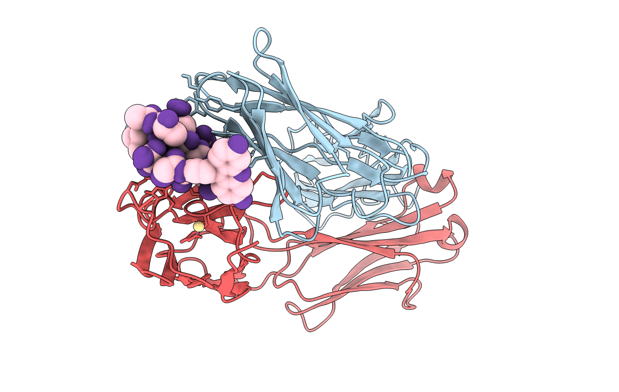
BACTERICIDAL ANTIBODY AGAINST NEISSERIA MENINGITIDIS
Biological Source:
Source Organism:
Mus musculus (Taxon ID: 10090)
Host Organism:
Method Details:
Experimental Method:
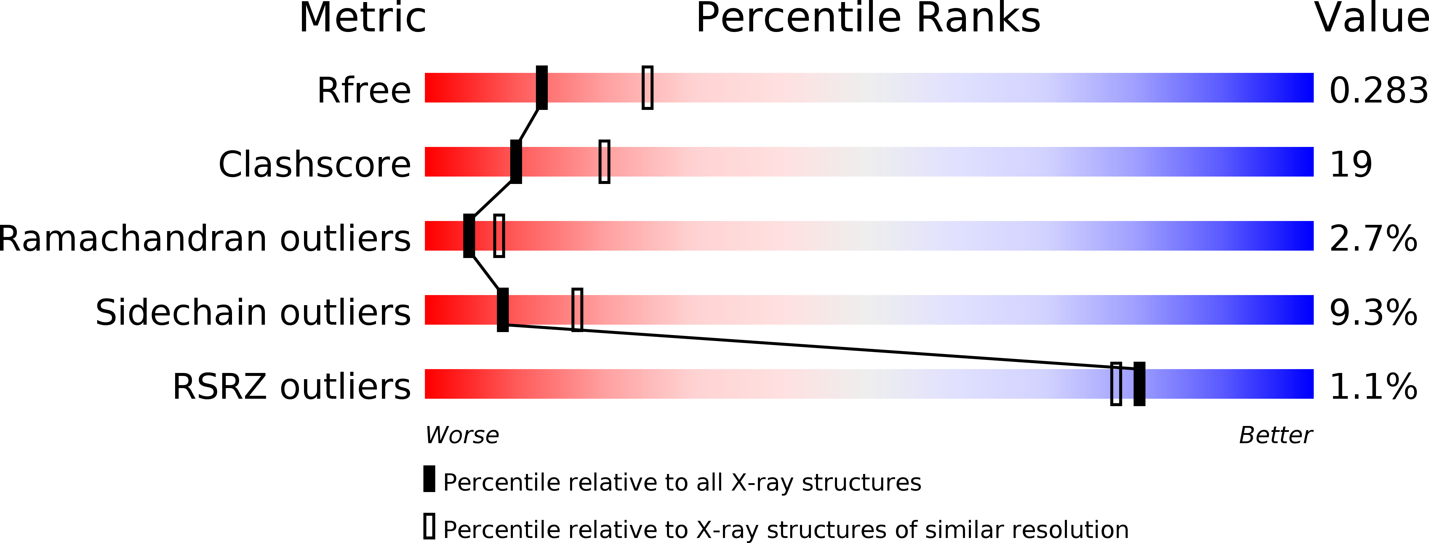
Resolution:
2.60 Å
R-Value Free:
0.30
R-Value Work:
0.19
R-Value Observed:
0.19
Space Group:
P 1 21 1
Macromolecular Entities
Structures with similar UniProt ID
Protein Blast
Polymer Type:polypeptide(L)
Molecule:MN12H2 IGG2A-KAPPA
Chain IDs:B (auth: H)
Chain Length:225
Number of Molecules:1
Biological Source:Mus musculus
Structures with similar UniProt ID
Protein Blast
Polymer Type:polypeptide(L)
Molecule:MN12H2 IGG2A-KAPPA
Chain IDs:A (auth: L)
Chain Length:219
Number of Molecules:1
Biological Source:Mus musculus
Modified Residue
Compound ID Chain ID Parent Comp ID Details 2D Image
CYF C CYS ?
THC C THR N-METHYLCARBONYLTHREONINE
Ligand Molecules
Primary Citation
Bactericidal antibody recognition of a PorA epitope of Neisseria meningitidis: crystal structure of a Fab fragment in complex with a fluorescein-conjugated peptide.
Proteins 29 113 125 (1997)
PMID: 9294871 DOI: 10.1002/(SICI)1097-0134(199709)29:1<113::AID-PROT9>3.3.CO;2-U

Abstact

Class 1 outer membrane protein PorA of Neisseria meningitidis is a vaccine candidate against bacterial meningitis. Antibodies against PorA are able to induce complement-mediated bacterial killing and thereby play an important role in protection against meningococcal disease. Bactericidal antibodies are all directed against variable regions VR1 and VR2 of the PorA sequence, corresponding to loops 1 and 4 of a two-dimensional topology model of the porin with eight extracellular loops. We have determined the crystal structure to 2.6 A resolution of the Fab fragment of bactericidal antibody MN12H2 against meningococcal PorA in complex with a linear fluorescein-conjugated peptide TKDTNNNL derived from the VR2 sequence of sero-subtype P1.7,16 (residues 180-187) from meningococcal strain H44/76. The peptide folds deeply into the binding cavity of the Fab molecule in a type I beta-turn, with the minimal P1.16 epitope DTNNN virtually completely buried. The structure reveals H-bonds and van der Waals interactions with all minimal epitope residues and one essential salt bridge between Asp-182 of the peptide and His-31 of the MN12H2 light chain. The key components of the recognition of PorA epitope P1.16 by bactericidal antibody MN12H2 correspond well with available thermodynamic data from binding studies. Furthermore, they indicate the structural basis of an increased endemic incidence of infection by group B meningococci in England and Wales since 1981 associated with the occurrence of an Neisseria meningitidis escape mutant (strain-MC58). The observed three-dimensional conformation of the peptide provides a rationale for the development of a synthetic peptide vaccine against meningococcal disease.

Legend

Protein

Chemical

Disease

Primary Citation of related structures