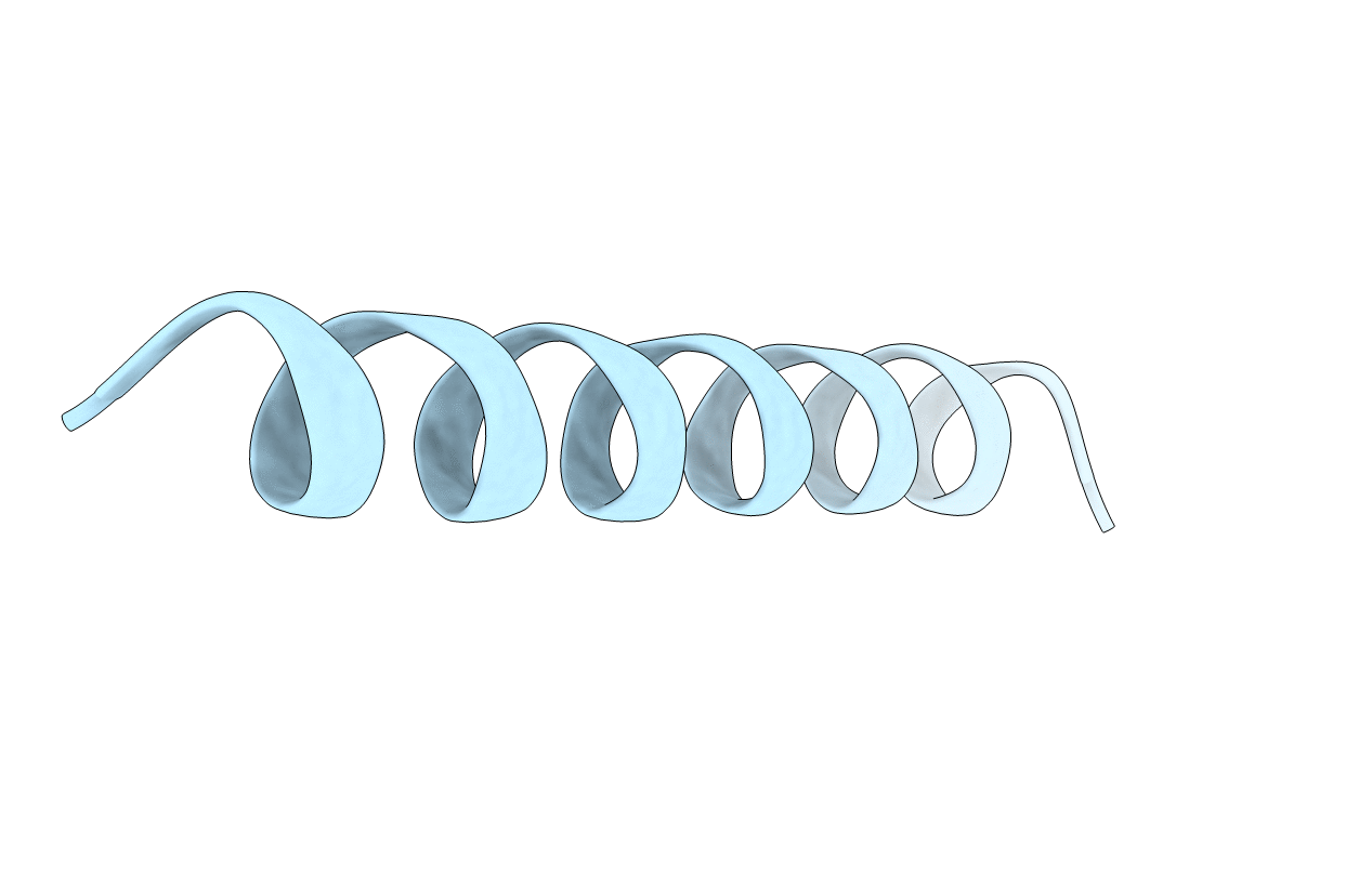
1MP6 image
Deposition Date 2002-09-11
Release Date 2002-09-25
Last Version Date 2024-05-22
Entry Detail
PDB ID:
1MP6
Title:
Structure of the transmembrane region of the M2 protein H+ channel by solid state NMR spectroscopy
Biological Source:
Source Organism:
(Taxon ID: )
Method Details:
Experimental Method:
Conformers Calculated:
30
Conformers Submitted:
1
Selection Criteria:
The lowest energy conformer with backbone and C beta atoms is deposited, preferred rotameric states of side chains were used during the backbone structure refinement but the side chain atoms were not included in the pdb file.
Macromolecular Entities
Polymer Type:polypeptide(L)
Molecule:Matrix protein M2
Chain IDs:A
Chain Length:25
Number of Molecules:1
Biological Source:
Ligand Molecules
Primary Citation
Structure of the transmembrane region of the M2 protein H(+) channel.
Protein Sci. 10 2241 2250 (2001)
PMID: 11604531 DOI: 10.1110/ps.17901

Abstact

The transmembrane domain of the M2 protein from influenza A virus forms a nearly uniform and ideal helix in a liquid crystalline bilayer environment. The exposure of the hydrophilic backbone structure is minimized through uniform hydrogen bond geometry imposed by the low dielectric lipid environment. A high-resolution structure of the monomer backbone and a detailed description of its orientation with respect to the bilayer were achieved using orientational restraints from solid-state NMR. With this unique information, the tetrameric structure of this H(+) channel is constrained substantially. Features of numerous published models are discussed in light of the experimental structure of the monomer and derived features of the tetrameric bundle.

Legend

Protein

Chemical

Disease

Primary Citation of related structures