Deposition Date
1999-04-30
Release Date
1999-05-06
Last Version Date
2024-10-16
Entry Detail
PDB ID:
1MNU
Keywords:
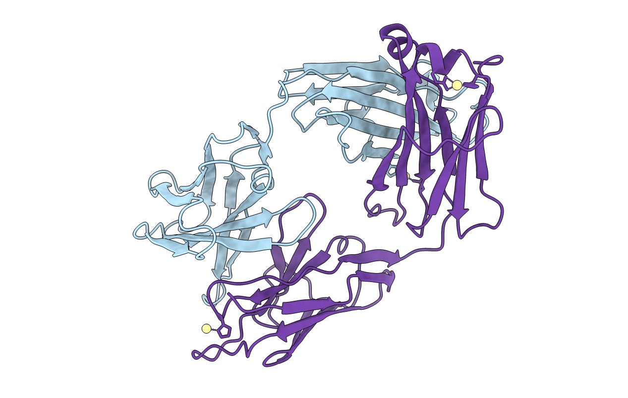
Title:
UNLIGANDED BACTERICIDAL ANTIBODY AGAINST NEISSERIA MENINGITIDIS
Biological Source:
Source Organism(s):
Mus musculus (Taxon ID: 10090)
Method Details:
Experimental Method:
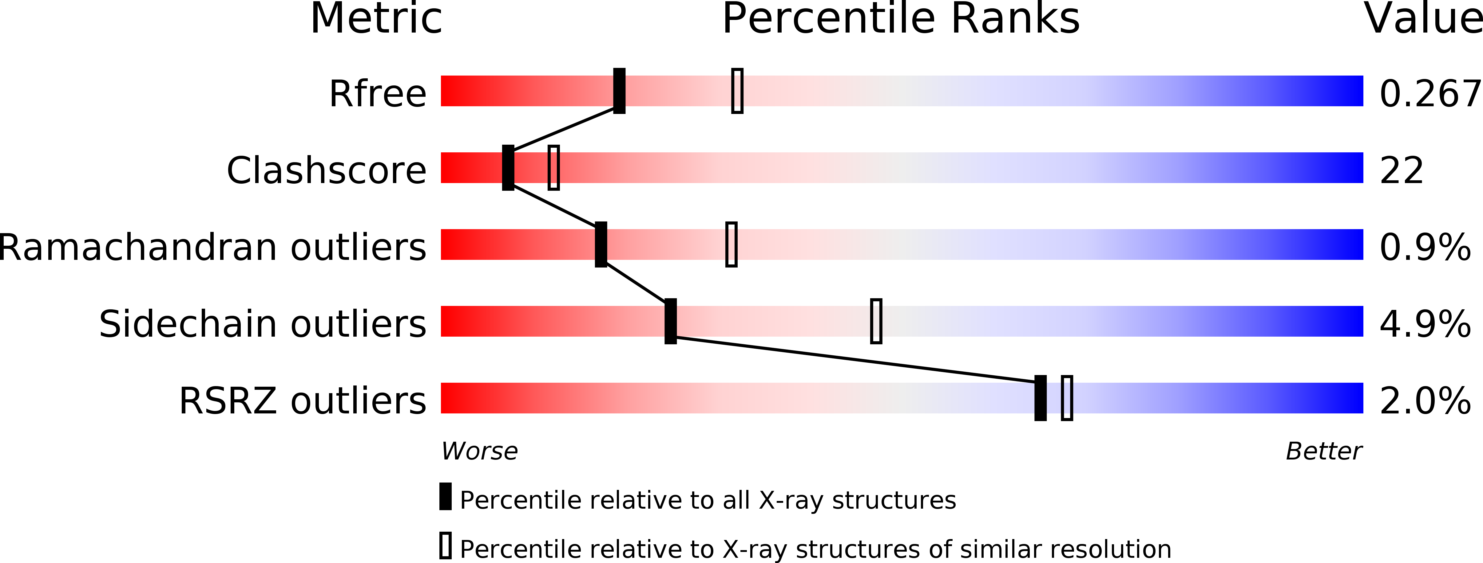
Resolution:
2.50 Å
R-Value Free:
0.27
R-Value Work:
0.23
Space Group:
C 1 2 1