1MMT image
Deposition Date 2002-09-04
Release Date 2003-09-04
Last Version Date 2023-11-15
Entry Detail
PDB ID:
1MMT
Keywords:
Title:
Crystal structure of ternary complex of the catalytic domain of human phenylalanine hydroxylase (Fe(II)) complexed with tetrahydrobiopterin and norleucine
Biological Source:
Source Organism:
Homo sapiens (Taxon ID: 9606)
Host Organism:
Method Details:
Experimental Method:
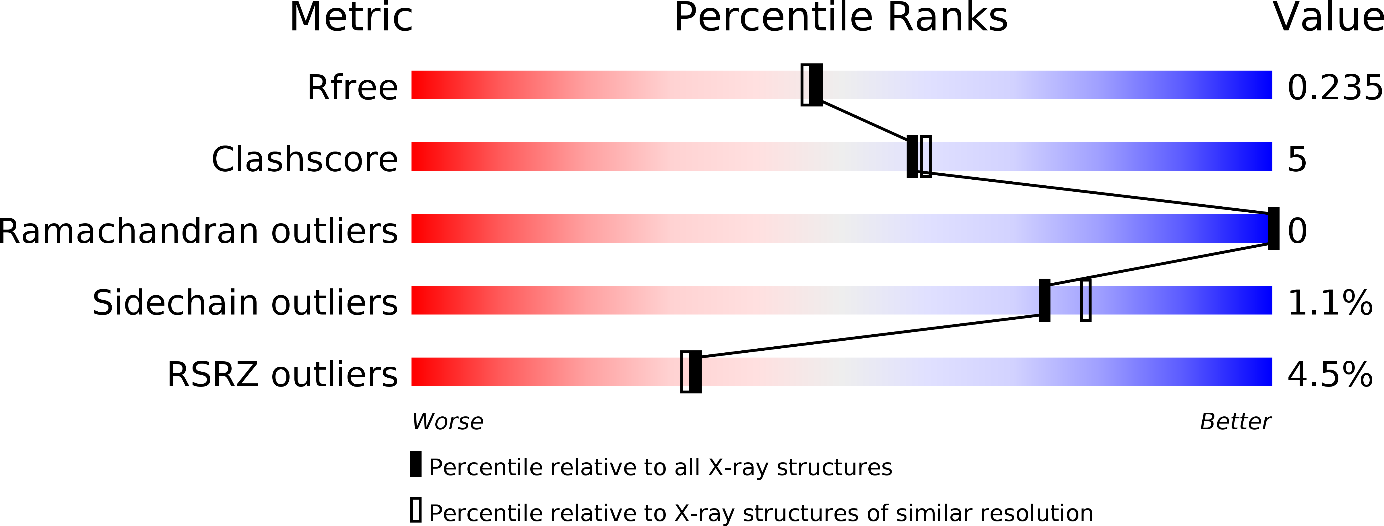
Resolution:
2.00 Å
R-Value Free:
0.24
R-Value Work:
0.21
Space Group:
C 2 2 21
Macromolecular Entities
Polymer Type:polypeptide(L)
Molecule:Phenylalanine-4-hydroxylase
Gene (Uniprot):PAH
Chain IDs:A
Chain Length:325
Number of Molecules:1
Biological Source:Homo sapiens
Primary Citation
2.0A resolution crystal structures of the ternary complexes of human phenylalanine hydroxylase catalytic domain with tetrahydrobiopterin and 3-(2-thienyl)-L-alanine or L-norleucine: substrate specificity and molecular motions related to substrate binding
J.Mol.Biol. 333 747 757 (2003)
PMID: 14568534 DOI: 10.1016/j.jmb.2003.09.004

Abstact

The crystal structures of the catalytic domain of human phenylalanine hydroxylase (hPheOH) in complex with the physiological cofactor 6(R)-L-erythro-5,6,7,8-tetrahydrobiopterin (BH(4)) and the substrate analogues 3-(2-thienyl)-L-alanine (THA) or L-norleucine (NLE) have been determined at 2.0A resolution. The ternary THA complex confirms a previous 2.5A structure, and the ternary NLE complex shows that similar large conformational changes occur on binding of NLE as those observed for THA. Both structures demonstrate that substrate binding triggers structural changes throughout the entire protomer, including the displacement of Tyr138 from a surface position to a buried position at the active site, with a maximum displacement of 20.7A for its hydroxyl group. Two hinge-bending regions, centred at Leu197 and Asn223, act in consort upon substrate binding to create further large structural changes for parts of the C terminus. Thus, THA/L-Phe binding to the active site is likely to represent the epicentre of the global conformational changes observed in the full-length tetrameric enzyme. The carboxyl and amino groups of THA and NLE are positioned identically in the two structures, supporting the conclusion that these groups are of key importance in substrate binding, thus explaining the broad non-physiological substrate specificity observed for artificially activated forms of the enzyme. However, the specific activity with NLE as the substrate was only about 5% of that with THA, which is explained by the different affinities of binding and different catalytic turnover.

Legend

Protein

Chemical

Disease

Primary Citation of related structures