1MMF image
Deposition Date 2002-09-03
Release Date 2003-06-10
Last Version Date 2024-02-14
Entry Detail
PDB ID:
1MMF
Keywords:
Title:
Crystal structure of substrate free form of glycerol dehydratase
Biological Source:
Source Organism:
Method Details:
Experimental Method:
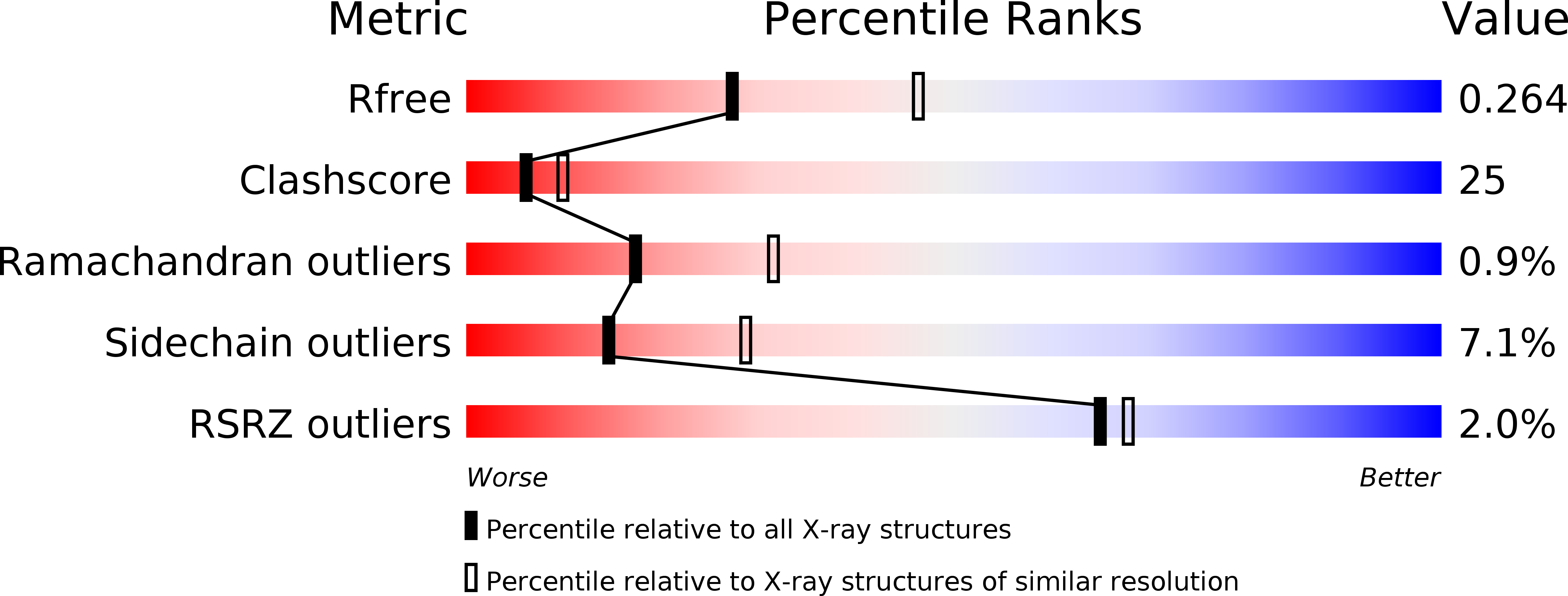
Resolution:
2.50 Å
R-Value Free:
0.26
R-Value Work:
0.22
Space Group:
P 1 21 1
Macromolecular Entities
Structures with similar UniProt ID
Protein Blast
Polymer Type:polypeptide(L)
Molecule:glycerol dehydrase alpha subunit
Gene (Uniprot):dhaB1
Chain IDs:A, D (auth: L)
Chain Length:555
Number of Molecules:2
Biological Source:Klebsiella pneumoniae
Structures with similar UniProt ID
Protein Blast
Polymer Type:polypeptide(L)
Molecule:glycerol dehydrase beta subunit
Gene (Uniprot):gldB
Chain IDs:B, E
Chain Length:194
Number of Molecules:2
Biological Source:Klebsiella pneumoniae
Structures with similar UniProt ID
Protein Blast
Polymer Type:polypeptide(L)
Molecule:glycerol dehydrase gamma subunit
Gene (Uniprot):dhaB3
Chain IDs:C (auth: G), F (auth: M)
Chain Length:141
Number of Molecules:2
Biological Source:Klebsiella pneumoniae
Primary Citation
Crystal structure of substrate free form of glycerol dehydratase
J.Inorg.Biochem. 93 84 91 (2003)
PMID: 12538056 DOI: 10.1016/S0162-0134(02)00523-8

Abstact

Glycerol dehydratase (GDH) and diol dehydratase (DDH) are highly homologous isofunctional enzymes that catalyze the elimination of water from glycerol and 1,2-propanediol (1,2-PD) to the corresponding aldehyde via a coenzyme B(12)-dependent radical mechanism. The crystal structure of substrate free form of GDH in complex with cobalamin and K(+) has been determined at 2.5 A resolution. Its overall fold and the subunit assembly closely resemble those of DDH. Comparison of this structure and the DDH structure, available only in substrate bound form, shows the expected change of the coordination of the essential K(+) from hexacoordinate to heptacoordinate with the displacement of a single coordinated water by the substrate diol. In addition, there appears to be an increase in the rigidity of the K(+) coordination (as measured by lower B values) upon the binding of the substrate. Structural analysis of the locations of conserved residues among various GDH and DDH sequences has aided in identification of residues potentially important for substrate preference or specificity of protein-protein interactions.

Legend

Protein

Chemical

Disease

Primary Citation of related structures