
Deposition Date
1995-03-21
Release Date
1996-08-17
Last Version Date
2024-02-14
Entry Detail
PDB ID:
1MMD
Keywords:
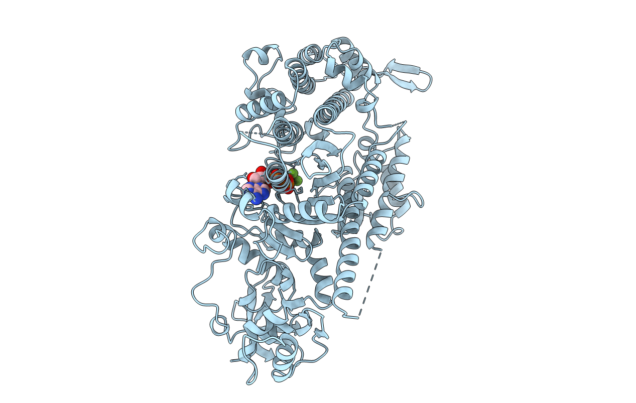
Title:
TRUNCATED HEAD OF MYOSIN FROM DICTYOSTELIUM DISCOIDEUM COMPLEXED WITH MGADP-BEF3
Biological Source:
Source Organism(s):
Dictyostelium discoideum (Taxon ID: 44689)
Expression System(s):
Method Details:
