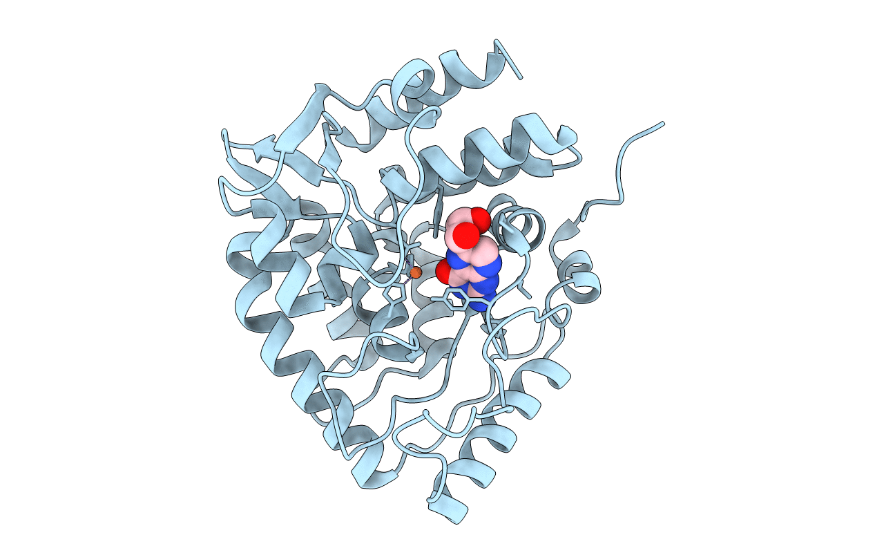
1MLW image
Deposition Date 2002-08-31
Release Date 2002-12-18
Last Version Date 2024-02-14
Entry Detail
PDB ID:
1MLW
Keywords:
Title:
Crystal structure of human tryptophan hydroxylase with bound 7,8-dihydro-L-biopterin cofactor and Fe(III)
Biological Source:
Source Organism:
Homo sapiens (Taxon ID: 9606)
Host Organism:
Method Details:
Experimental Method:
Resolution:
1.71 Å
R-Value Free:
0.22
R-Value Work:
0.20
Space Group:
P 21 21 21
Macromolecular Entities
Polymer Type:polypeptide(L)
Molecule:Tryptophan 5-monooxygenase
Gene (Uniprot):TPH1
Chain IDs:A
Chain Length:301
Number of Molecules:1
Biological Source:Homo sapiens
Primary Citation
Three-dimensional structure of human tryptophan hydroxylase and its implications for the biosynthesis of the neurotransmitters serotonin and melatonin
Biochemistry 41 12569 12574 (2002)
PMID: 12379098 DOI: 10.1021/bi026561f

Abstact

Tryptophan hydroxylase oxidizes L-tryptophan to 5-hydroxy-L-tryptophan in the rate-determining step of serotonin biosynthesis. We have determined the X-ray crystal structure (1.7 A) of a truncated functional form of human tryptophan hydroxylase with the bound cofactor analogue 7,8-dihydro-L-biopterin, providing the first atomic-resolution information for the catalytic domain of this important enzyme. Comparison of the three-dimensional structures of all three members of the aromatic amino acid hydroxylase family--tyrosine hydroxylase, phenylalanine hydroxylase, and tryptophan hydroxylase--reveals important differences at the active sites.

Legend

Protein

Chemical

Disease

Primary Citation of related structures