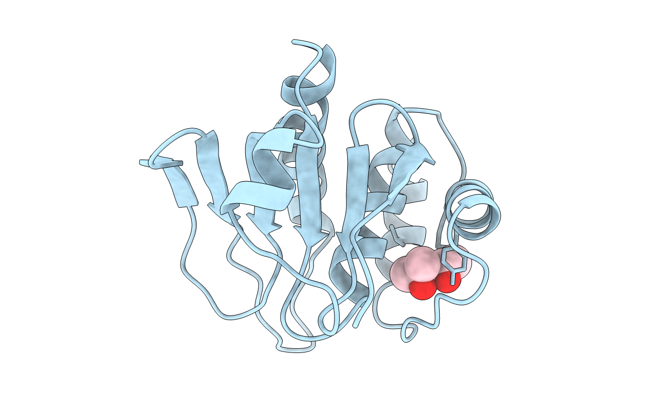
1MKP image
Deposition Date 1998-07-11
Release Date 1999-07-22
Last Version Date 2024-02-14
Entry Detail
PDB ID:
1MKP
Keywords:
Title:
CRYSTAL STRUCTURE OF PYST1 (MKP3)
Biological Source:
Source Organism:
Homo sapiens (Taxon ID: 9606)
Method Details:
Experimental Method:
Resolution:
2.35 Å
R-Value Free:
0.23
R-Value Work:
0.20
R-Value Observed:
0.20
Space Group:
P 41 21 2
Macromolecular Entities
Structures with similar UniProt ID
Protein Blast
Polymer Type:polypeptide(L)
Molecule:PYST1
Gene (Uniprot):DUSP6
Mutagens:C293S
Chain IDs:A
Chain Length:144
Number of Molecules:1
Biological Source:Homo sapiens
Primary Citation
Crystal structure of the MAPK phosphatase Pyst1 catalytic domain and implications for regulated activation.
Nat.Struct.Biol. 6 174 181 (1999)
PMID: 10048930 DOI: 10.1038/5861

Abstact

The crystal structure of the catalytic domain from the MAPK phosphatase Pyst1 (Pyst1-CD) has been determined at 2.35 A. The structure adopts a protein tyrosine phosphatase (PTPase) fold with a shallow active site that displays a distorted geometry in the absence of its substrate with some similarity to the dual-specificity phosphatase cdc25. Functional characterization of Pyst1-CD indicates it is sufficient to dephosphorylate activated ERK2 in vitro. Kinetic analysis of Pyst1 and Pyst1-CD using the substrate p-nitrophenyl phosphate (pNPP) reveals that both molecules undergo catalytic activation in the presence of recombinant inactive ERK2, switching from a low- to high-activity form. Mutation of Asp 262, located 5.5 A distal to the active site, demonstrates it is essential for catalysis in the high-activity ERK2-dependent conformation of Pyst1 but not for the low-activity ERK2-independent form, suggesting that ERK2 induces closure of the Asp 262 loop over the active site, thereby enhancing Pyst1 catalytic efficiency.

Legend

Protein

Chemical

Disease

Primary Citation of related structures