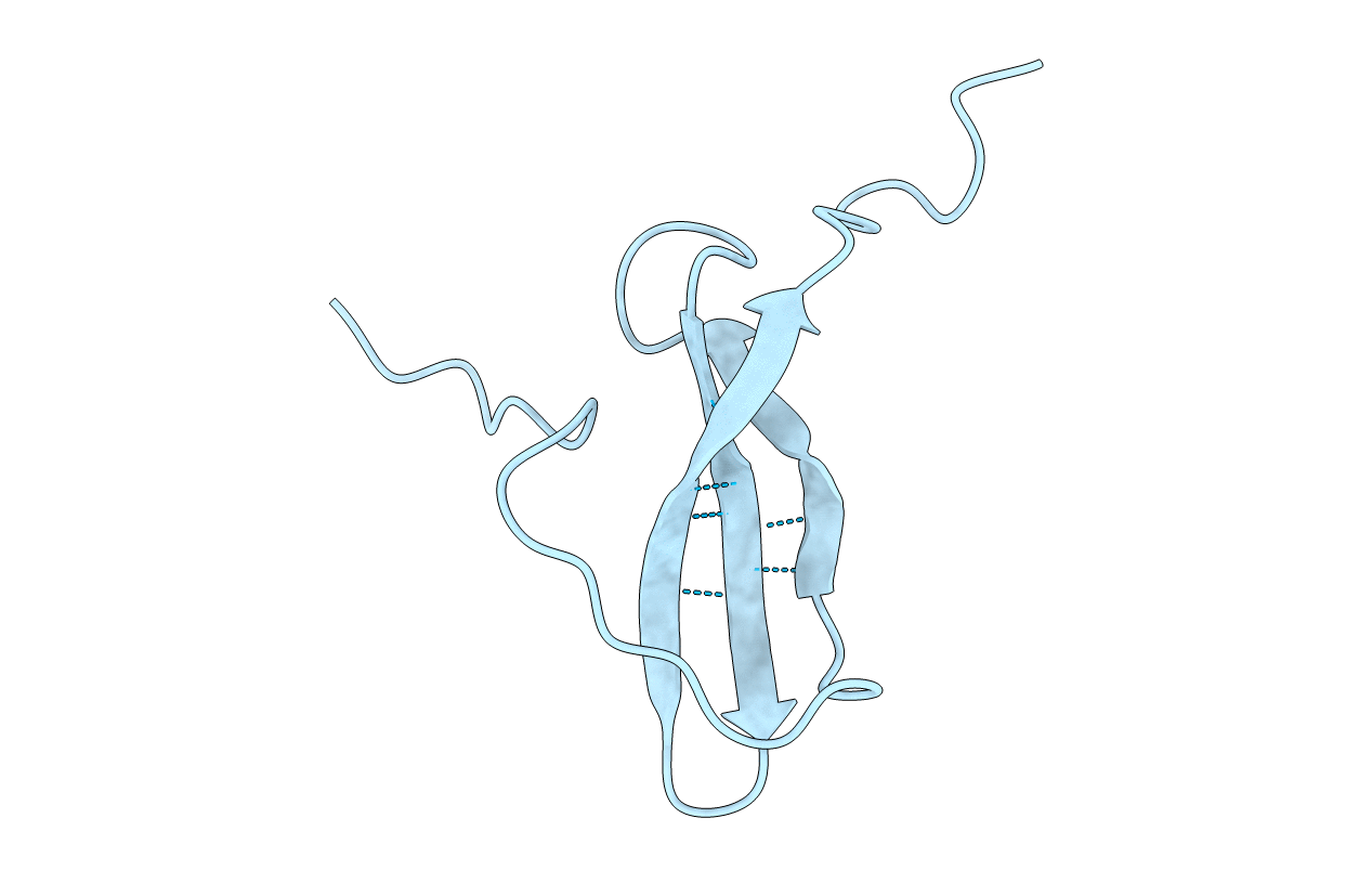
1MKN image
Entry Detail
PDB ID:
1MKN
Title:
N-TERMINAL HALF OF MIDKINE
Biological Source:
Source Organism:
PDB Version:
Deposition Date:
1999-03-16
Release Date:
1999-03-23
Method Details:
Experimental Method:
Conformers Submitted:
1
Macromolecular Entities
Polymer Type:polypeptide(L)
Description:PROTEIN (MIDKINE)
Chain IDs:A
Chain Length:59
Number of Molecules:1
Biological Source:
Ligand Molecules
Primary Citation
Solution structure of midkine, a new heparin-binding growth factor.
EMBO J. 16 6936 6946 (1997)
PMID: 9384573 DOI: 10.1093/emboj/16.23.6936

Abstact

Midkine (MK) is a 13 kDa heparin-binding polypeptide which enhances neurite outgrowth, neuronal cell survival and plasminogen activator activity. MK is structurally divided into two domains, and most of the biological activities are located on the C-terminal domain. The solution structures of the two domains were determined by NMR. Both domains consist of three antiparallel beta-strands, but the C-terminal domain has a long flexible hairpin loop where a heparin-binding consensus sequence is located. Basic residues on the beta-sheet of the C-terminal domain form another heparin-binding site. Measurement of NMR signals in the presence of a heparin oligosaccharides verified that multiple amino acids in the two sites participated in heparin binding. The MK dimer has been shown to be the active form, giving signals to endothelial cells and probably to neuronal cells. We present a head-to-head dimer model of MK. The model was supported by the results of cross-linking experiments using transglutaminase. The dimer has a fused heparin-binding site at the dimer interface of the C-terminal domain, and the heparin-binding sites on MK fit the sulfate group clusters on heparin. These features are consistent with the proposed stronger heparin-binding activity and biological activity of the dimer.

Legend

Protein

Chemical

Disease

Primary Citation of related structures