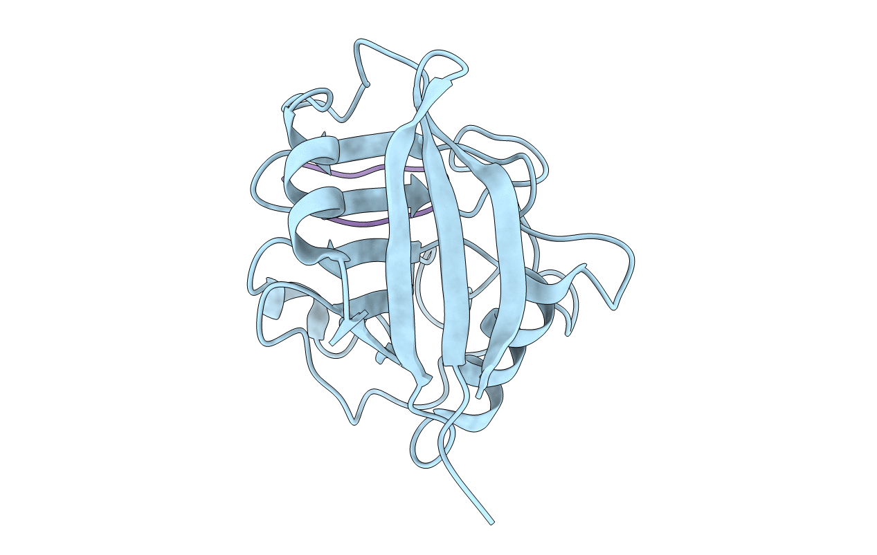
1MIK image
Deposition Date 1995-09-20
Release Date 1996-03-08
Last Version Date 2024-06-05
Entry Detail
PDB ID:
1MIK
Title:
THE ROLE OF WATER MOLECULES IN THE STRUCTURE-BASED DESIGN OF (5-HYDROXYNORVALINE)-2-CYCLOSPORIN: SYNTHESIS, BIOLOGICAL ACTIVITY, AND CRYSTALLOGRAPHIC ANALYSIS WITH CYCLOPHILIN A
Biological Source:
Source Organism:
Host Organism:
Method Details:
Experimental Method:
Resolution:
1.76 Å
R-Value Work:
0.17
R-Value Observed:
0.17
Space Group:
P 21 21 21
Macromolecular Entities
Polymer Type:polypeptide(L)
Molecule:PEPTIDYL-PROLYL CIS-TRANS ISOMERASE A
Chain IDs:A
Chain Length:165
Number of Molecules:1
Biological Source:HOMO SAPIENS
Polymer Type:polypeptide(L)
Molecule:CYCLOSPORIN A
Mutations:YES
Chain IDs:B
Chain Length:11
Number of Molecules:1
Biological Source:TOLYPOCLADIUM INFLATUM
Peptide-like Molecules
PRD_000766
Primary Citation
The Role of Water Molecules in the Structure-Based Design of (5-Hydroxynorvaline)-2-Cyclosporin: Synthesis, Biological Activity, and Crystallographic Analysis with Cyclophilin A.
J.Med.Chem. 38 3361 ? (1995)
PMID: 7650689 DOI: 10.1021/JM00017A020

Abstact

Analysis of the contact surface of the cyclophilin A (CypA)/cyclosporin A (CsA, 1) crystal structure delineates a unique cavity between both molecules in the vicinity of the Abu-2 side chain atoms of 1 (Abu pocket). Therefore, (5-hydroxynorvaline)-2-cyclosporin (2) was designed and prepared as a CsA derivative which could mediate additional interactions within the pocket. The X-ray crystal structure of the CypA/2 complex at 1.76 A resolution shows that 1 and 2 have identical backbone conformations and that the introduced hydroxypropyl chain makes indeed the expected supplemental interactions with CypA. However, 2 has 8-9-fold lower binding affinity than 1 for CypA. This results from a presumed unfavorable free energy change associated with the displacement of one of the tightly bound water molecules within the pocket and a change in prebinding equilibria. The role of the later was assessed by comparing the conformation distribution of 1 and 2 to that of norvaline-2-cyclosporin (3) and norvaline-2-(D-MeSer)-3-cyclosporin (4).

Legend

Protein

Chemical

Disease