1MI7 image
Deposition Date 2002-08-22
Release Date 2003-09-02
Last Version Date 2024-02-14
Entry Detail
PDB ID:
1MI7
Keywords:
Title:
Crystal Structure of Domain Swapped trp Aporepressor in 30%(v/v) Isopropanol
Biological Source:
Source Organism:
Escherichia coli (Taxon ID: 562)
Method Details:
Experimental Method:
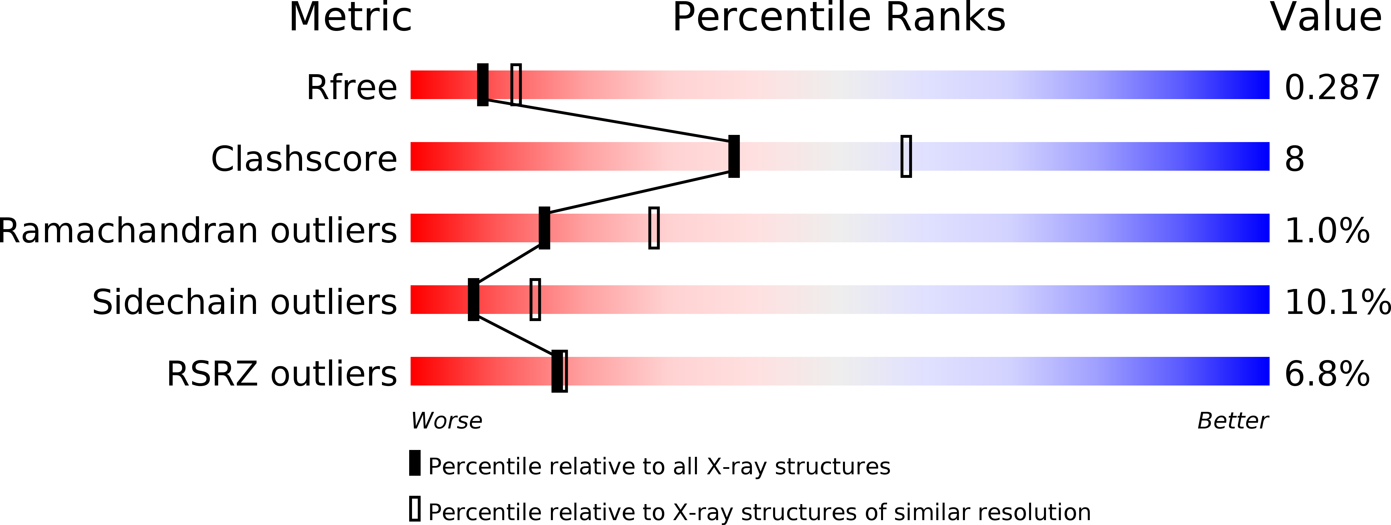
Resolution:
2.50 Å
R-Value Free:
0.28
R-Value Work:
0.25
R-Value Observed:
0.25
Space Group:
P 61 2 2
Macromolecular Entities
Polymer Type:polypeptide(L)
Molecule:Trp operon repressor
Gene (Uniprot):trpR
Chain IDs:A (auth: R)
Chain Length:107
Number of Molecules:1
Biological Source:Escherichia coli
Ligand Molecules
Primary Citation
E. coli trp repressor forms a domain-swapped array in aqueous alcohol.
Structure 12 1099 1108 (2004)
PMID: 15274929 DOI: 10.1016/j.str.2004.03.019

Abstact

The E. coli trp repressor (trpR) homodimer recognizes its palindromic DNA binding site through a pair of flexible helix-turn-helix (HTH) motifs displayed on an intertwined helical core. Flexible N-terminal arms mediate association between dimers bound to tandem DNA sites. The 2.5 A X-ray structure of trpR crystallized in 30% (v/v) isopropanol reveals a substantial conformational rearrangement of HTH motifs and N-terminal arms, with the protein appearing in the unusual form of an ordered 3D domain-swapped supramolecular array. Small angle X-ray scattering measurements show that the self-association properties of trpR in solution are fundamentally altered by isopropanol.

Legend

Protein

Chemical

Disease

Primary Citation of related structures