Deposition Date
2002-08-21
Release Date
2003-08-05
Last Version Date
2024-04-03
Entry Detail
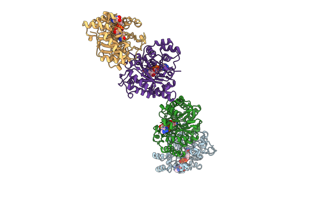
PDB ID:
1MI3
Keywords:
Title:
1.8 Angstrom structure of xylose reductase from Candida tenuis in complex with NAD
Biological Source:
Source Organism(s):
Candida tenuis (Taxon ID: 45596)
Expression System(s):
Method Details:
Experimental Method:
Resolution:
1.80 Å
R-Value Free:
0.21
R-Value Work:
0.18
R-Value Observed:
0.18
Space Group:
C 1 2 1