Deposition Date
2002-08-21
Release Date
2002-09-18
Last Version Date
2024-02-14
Entry Detail
PDB ID:
1MHX
Keywords:
Title:
Crystal Structures of the redesigned protein G variant NuG1
Biological Source:
Source Organism(s):
Finegoldia magna (Taxon ID: 334413)
Expression System(s):
Method Details:
Experimental Method:
Resolution:
1.80 Å
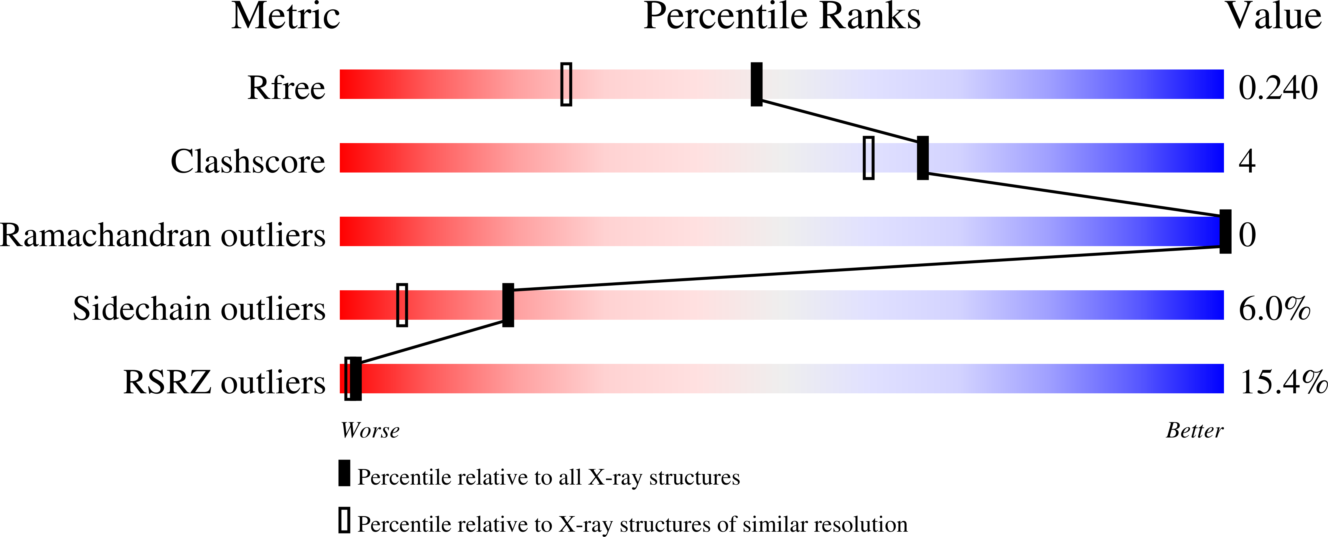
R-Value Free:
0.22
R-Value Work:
0.21
R-Value Observed:
0.26
Space Group:
I 4 2 2