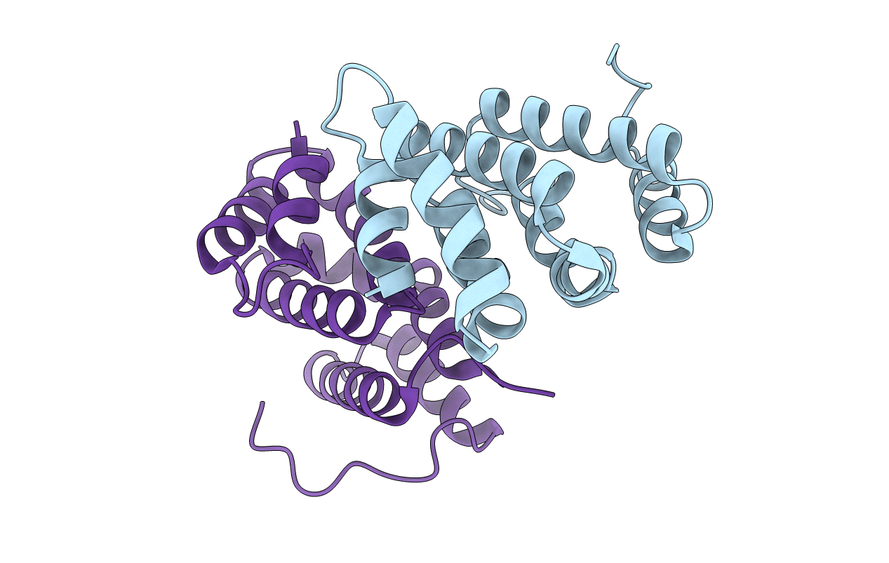
1MHQ image
Entry Detail
PDB ID:
1MHQ
Title:
Crystal Structure Of Human GGA2 VHS Domain
Biological Source:
Source Organism:
Host Organism:
PDB Version:
Deposition Date:
2002-08-20
Release Date:
2003-03-11
Method Details:
Experimental Method:
Resolution:
2.20 Å
R-Value Free:
0.26
R-Value Work:
0.22
R-Value Observed:
0.22
Space Group:
P 21 21 21
Macromolecular Entities
Polymer Type:polypeptide(L)
Description:ADP-ribosylation factor binding protein GGA2
Chain IDs:A, B
Chain Length:148
Number of Molecules:2
Biological Source:Homo sapiens
Primary Citation
Crystal structure of GGA2 VHS domain and its implication in plasticity in the ligand binding pocket
FEBS LETT. 537 171 176 (2003)
PMID: 12606052 DOI: 10.1016/S0014-5793(03)00095-4

Abstact

Golgi-localized, gamma-ear-containing, ARF binding (GGA) proteins regulate intracellular vesicle transport by recognizing sorting signals on the cargo surface in the initial step of the budding process. The VHS (VPS27, Hrs, and STAM) domain of GGA binds with the signal peptides. Here, a crystal structure of the VHS domain of GGA2 is reported at 2.2 A resolution, which permits a direct comparison with that of homologous proteins, GGA1 and GGA3. Significant structural difference is present in the loop between helices 6 and 7, which forms part of the ligand binding pocket. Intrinsic fluorescence spectroscopic study indicates that this loop undergoes a conformational change upon ligand binding. Thus, the current structure suggests that a conformational change induced by ligand binding occurs in this part of the ligand pocket.

Legend

Protein

Chemical

Disease

Primary Citation of related structures