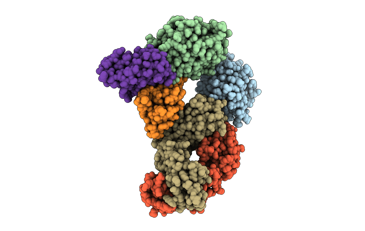
1MHP image
Deposition Date 2002-08-20
Release Date 2003-04-15
Last Version Date 2024-11-06
Entry Detail
PDB ID:
1MHP
Keywords:
Title:
Crystal structure of a chimeric alpha1 integrin I-domain in complex with the Fab fragment of a humanized neutralizing antibody
Biological Source:
Source Organism:
Rattus norvegicus (Taxon ID: 10116)
Mus musculus (Taxon ID: 10090)
Host Organism:
Method Details:
Experimental Method:
Resolution:
2.80 Å
R-Value Free:
0.27
R-Value Work:
0.21
R-Value Observed:
0.22
Space Group:
P 65
Macromolecular Entities
Polymer Type:polypeptide(L)
Molecule:integrin alpha 1, (RESIDUES 169-360)
Gene (Uniprot):Itga1
Mutations:G217V, R218Q, Q219R, L222R
Chain IDs:A, D (auth: B)
Chain Length:192
Number of Molecules:2
Biological Source:Rattus norvegicus
Polymer Type:polypeptide(L)
Molecule:Fab fragment, heavy chain
Chain IDs:B (auth: H), E (auth: X)
Chain Length:219
Number of Molecules:2
Biological Source:Mus musculus
Polymer Type:polypeptide(L)
Molecule:FAB FRAGMENT, light chain
Chain IDs:C (auth: L), F (auth: Y)
Chain Length:212
Number of Molecules:2
Biological Source:Mus musculus
Ligand Molecules
Primary Citation
Crystal Structure of the alpha 1 beta 1 Integrin I Domain in Complex with an Antibody Fab Fragment
J.Mol.Biol. 327 1031 1041 (2003)
PMID: 12662928 DOI: 10.1016/S0022-2836(03)00203-1

Abstact

The alpha1beta1 (VLA-1) integrin is a cell-surface receptor for collagen and laminin and has been implicated in biological pathways involved in several pathological processes. These processes may be inhibited by the monoclonal antibody AQC2, which binds with high affinity to human alpha1beta1 integrin. To understand the structural basis of the inhibition we determined the crystal structure of the complex of a chimeric rat/human I domain of the alpha1beta1 integrin and the Fab fragment of humanized AQC2 antibody. The structure of the complex shows that the antibody blocks the collagen binding site of the I domain. An aspartate residue, from the CDR3 loop of the antibody heavy chain, coordinates the MIDAS metal ion in a manner similar to that of a glutamate residue from collagen. Substitution of the aspartate residue by alanine or arginine results in significant reduction of antibody binding affinity. Interestingly, although the mode of metal ion coordination resembles that of the open conformation, the I domain maintains an overall closed conformation previously observed only for unliganded I domains.

Legend

Protein

Chemical

Disease

Primary Citation of related structures