1MH0 image
Deposition Date 2002-08-18
Release Date 2002-11-08
Last Version Date 2024-11-13
Entry Detail
PDB ID:
1MH0
Keywords:
Title:
Crystal structure of the anticoagulant slow form of thrombin
Biological Source:
Source Organism:
Homo sapiens (Taxon ID: 9606)
Host Organism:
Method Details:
Experimental Method:
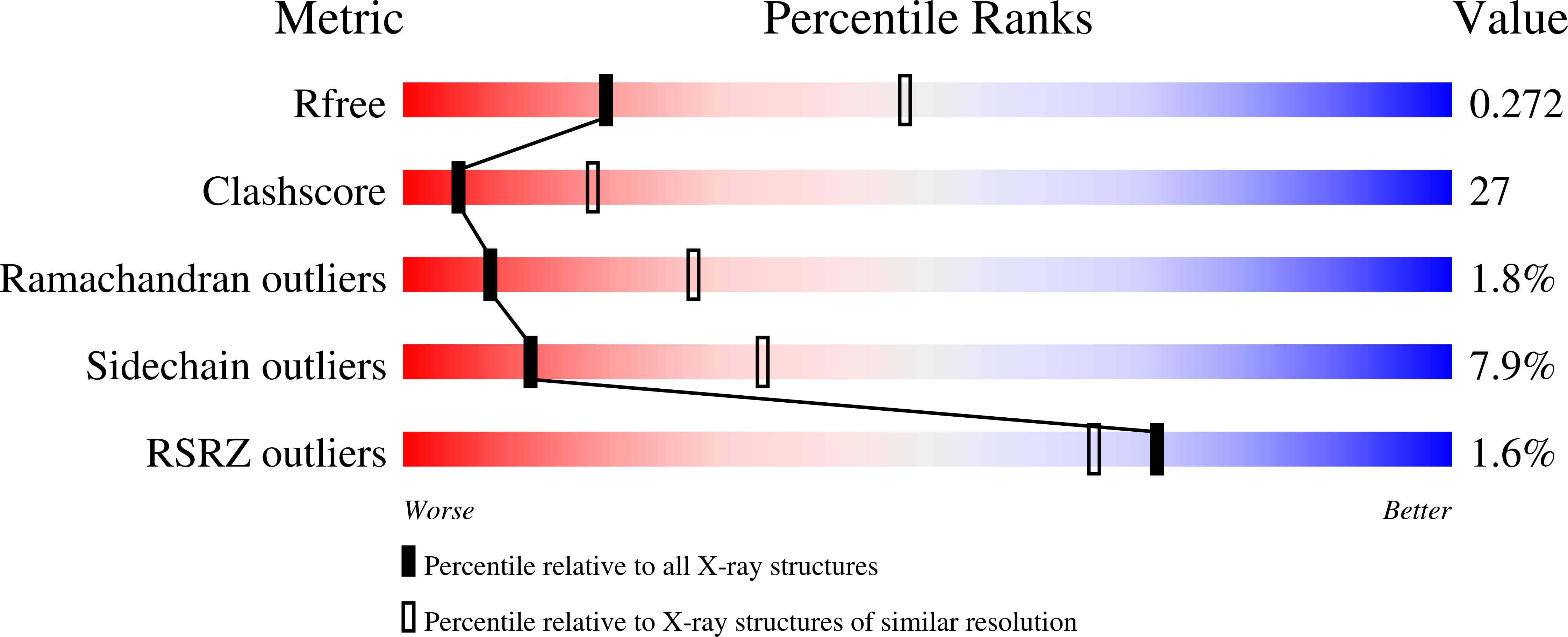
Resolution:
2.80 Å
R-Value Free:
0.27
R-Value Work:
0.21
R-Value Observed:
0.21
Space Group:
P 21 21 21
Macromolecular Entities
Polymer Type:polypeptide(L)
Molecule:Prothrombin
Gene (Uniprot):F2
Mutations:R77aA
Chain IDs:A, B
Chain Length:287
Number of Molecules:2
Biological Source:Homo sapiens
Ligand Molecules
Primary Citation
Crystal structure of the anticoagulant slow form of thrombin
J.Biol.Chem. 277 40177 40180 (2002)
PMID: 12205081 DOI: 10.1074/jbc.C200465200

Abstact

Using the thrombin mutant R77aA devoid of the site of autoproteolytic degradation at exosite I, we have solved for the first time the structure of thrombin free of any inhibitors and effector molecules and stabilized in the Na(+)-free slow form. The slow form shows subtle differences compared with the currently available structures of the Na(+)-bound fast form that carry inhibitors at the active site or exosite I. The most notable differences are the displacement of Asp-189 in the S1 specificity pocket, a downward shift of the 190-193 strand, a rearrangement of the side chain of Glu-192, and a significant shift in the position of the catalytic Ser-195 that is no longer within H-bonding distance from His-57. The structure of the slow form explains the reduced specificity toward synthetic and natural substrates and suggests a molecular basis for its anticoagulant properties.

Legend

Protein

Chemical

Disease

Primary Citation of related structures