1MG5 image
Deposition Date 2002-08-14
Release Date 2003-10-14
Last Version Date 2024-02-14
Entry Detail
PDB ID:
1MG5
Keywords:
Title:
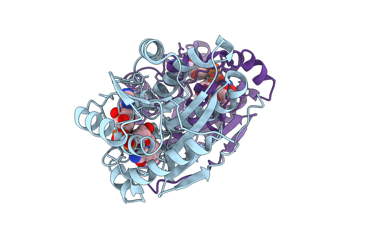
Crystal structure of Drosophila melanogaster alcohol dehydrogenase complexed with NADH and acetate at 1.6 A
Biological Source:
Source Organism:
Method Details:
Experimental Method:
Resolution:
1.63 Å
R-Value Free:
0.20
R-Value Work:
0.16
Space Group:
P 1 21 1
Macromolecular Entities
Polymer Type:polypeptide(L)
Molecule:alcohol dehydrogenase
Gene (Uniprot):Adh
Chain IDs:A, B
Chain Length:255
Number of Molecules:2
Biological Source:Drosophila melanogaster
Primary Citation
Drosophila alcohol dehydrogenase: acetate-enzyme interactions and novel insights into the effects of electrostatics on catalysis
J.Mol.Biol. 345 579 598 (2005)
PMID: 15581900 DOI: 10.1016/j.jmb.2004.10.028

Abstact

Drosophila alcohol dehydrogenase (DADH) is an NAD+-dependent enzyme that catalyzes the oxidation of alcohols to aldehydes/ketones and that is also able to further oxidize aldehydes to their corresponding carboxylic acids. The structure of the ternary enzyme-NADH-acetate complex of the slow alleloform of Drosophila melanogaster ADH (DmADH-S) was solved at 1.6 A resolution by X-ray crystallography. The coenzyme stereochemistry of the aldehyde dismutation reaction showed that the obtained enzyme-NADH-acetate complex reflects a productive ternary complex although no enzymatic reaction occurs. The stereochemistry of the acetate binding in the bifurcated substrate-binding site, along with previous stereochemical studies of aldehyde reduction and alcohol oxidation shows that the methyl group of the aldehyde in the reduction reaction binds to the R1 and in the oxidation reaction to the R2 sub-site. NMR studies along with previous kinetic studies show that the formed acetaldehyde intermediate in the oxidation of ethanol to acetate leaves the substrate site prior to the reduced coenzyme, and then binds to the newly formed enzyme-NAD+ complex. Here, we compare the three-dimensional structure of D.melanogaster ADH-S and a previous theoretically built model, evaluate the differences with the crystal structures of five Drosophila lebanonensis ADHs in numerous complexed forms that explain the substrate specificity as well as subtle kinetic differences between these two enzymes based on their crystal structures. We also re-examine the electrostatic influence of charged residues on the surface of the protein on the catalytic efficiency of the enzyme.

Legend

Protein

Chemical

Disease

Primary Citation of related structures