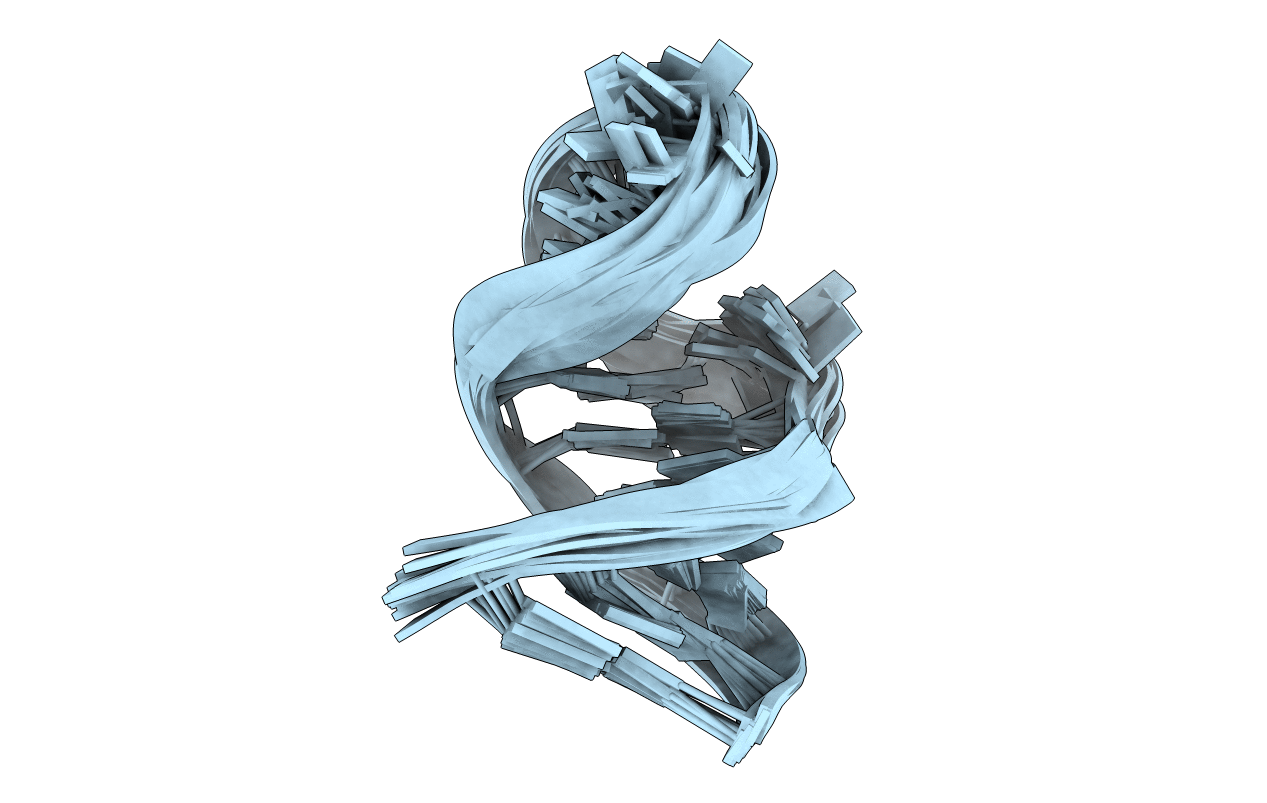
1MFK image
Deposition Date 2002-08-12
Release Date 2002-11-13
Last Version Date 2024-05-22
Entry Detail
PDB ID:
1MFK
Keywords:
Title:
Structure of Prokaryotic SECIS mRNA Hairpin
Biological Source:
Source Organism:
(Taxon ID: )
Method Details:
Experimental Method:
Conformers Calculated:
200
Conformers Submitted:
20
Selection Criteria:
structures with the lowest energy
Macromolecular Entities
Polymer Type:polyribonucleotide
Molecule:5'-R(P*GP*GP*CP*GP*GP*UP*UP*GP*CP*AP*GP*GP*UP*CP*UP*GP*CP*AP*CP*CP*GP*CP*C)-3'
Chain IDs:A
Chain Length:23
Number of Molecules:1
Biological Source:
Ligand Molecules
Primary Citation
Structure of Prokaryotic SECIS mRNA Hairpin and its Interaction with Elongation Factor SELB
J.Mol.Biol. 324 137 150 (2002)
PMID: 12421564 DOI: 10.1016/S0022-2836(02)01030-6

Abstact

In prokaryotes, the recoding of a UGA stop codon as a selenocysteine codon requires a special elongation factor (EF) SelB and a stem-loop structure within the mRNA called a selenocysteine insertion sequence (SECIS). Here, we used NMR spectroscopy to determine the solution structure of the SECIS mRNA hairpin and characterized its interaction with the mRNA-binding domain of SelB. Our structural and biochemical data identified the conserved structural features important for binding to EF SelB within different SECIS RNA sequences. In the free SECIS mRNA structure, conserved nucleotides are strongly exposed for recognition by SelB. Binding of the C-terminal domain of SelB stabilizes the RNA secondary structure. In the protein-RNA complex, a Watson-Crick loop base-pair leaves a GpU sequence accessible for SelB recognition. This GpU sequence at the tip of the capping tetraloop and a bulge uracil five Watson-Crick base-pairs apart from the GpU are essential for interaction with SelB.

Legend

Protein

Chemical

Disease

Primary Citation of related structures