Deposition Date
1994-06-09
Release Date
1994-08-31
Last Version Date
2024-02-14
Entry Detail
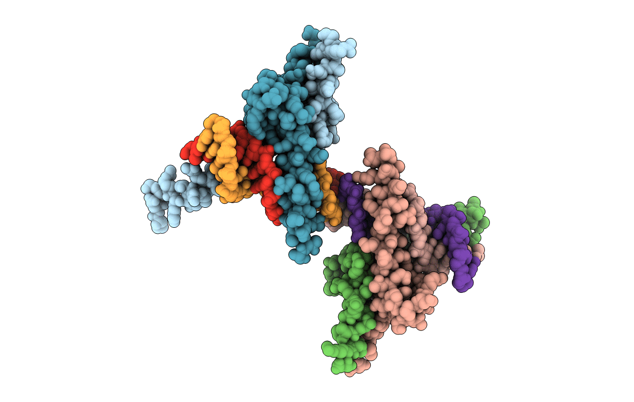
PDB ID:
1MDY
Keywords:
Title:
CRYSTAL STRUCTURE OF MYOD BHLH DOMAIN BOUND TO DNA: PERSPECTIVES ON DNA RECOGNITION AND IMPLICATIONS FOR TRANSCRIPTIONAL ACTIVATION
Biological Source:
Source Organism(s):
Mus musculus (Taxon ID: 10090)
Method Details:
Experimental Method:
Resolution:
2.80 Å
R-Value Free:
0.33
R-Value Work:
0.25
R-Value Observed:
0.25
Space Group:
P 21 21 2