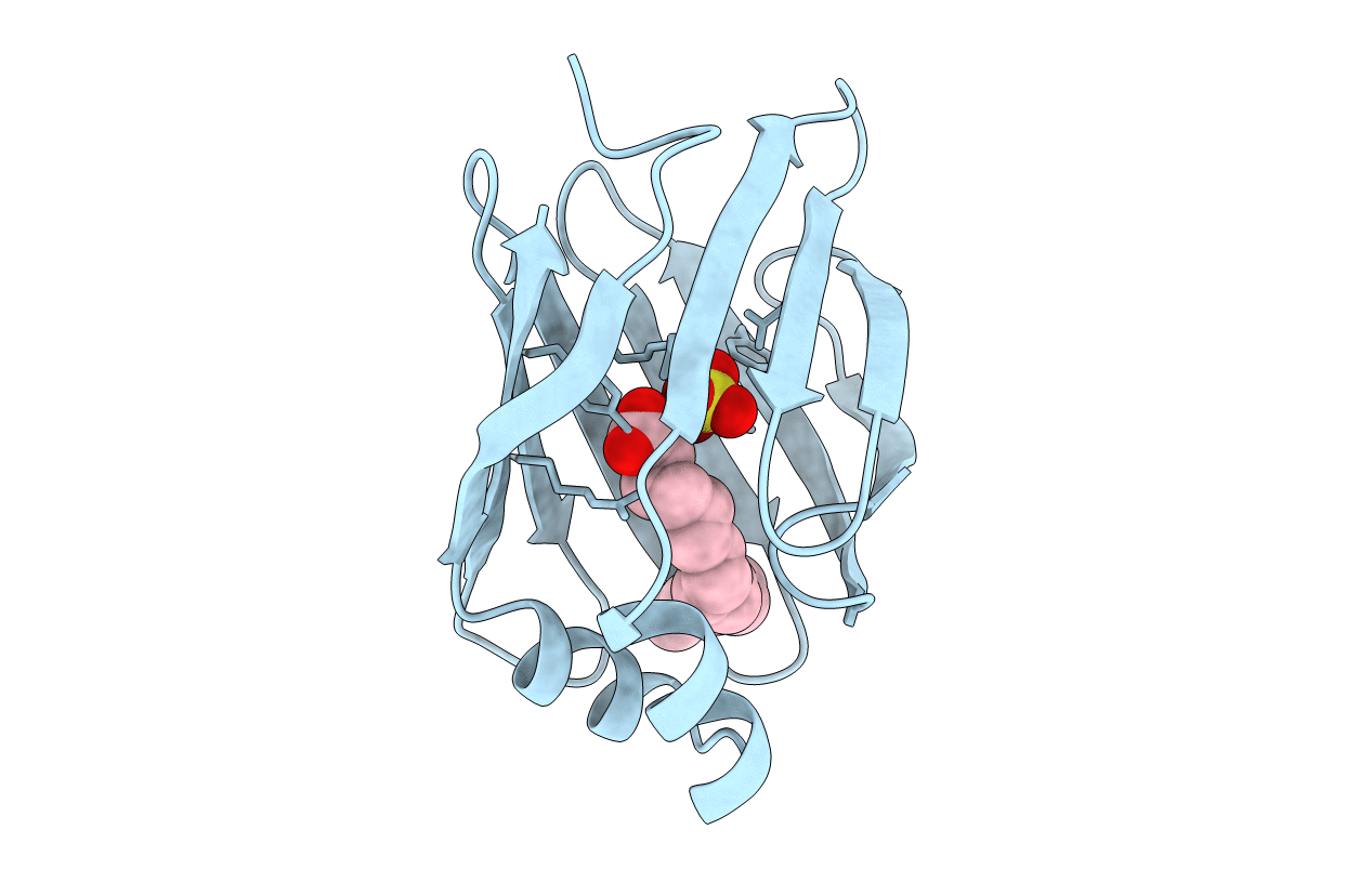
1MDC image
Deposition Date 1992-07-20
Release Date 1994-01-31
Last Version Date 2024-11-06
Entry Detail
PDB ID:
1MDC
Keywords:
Title:
CRYSTALLIZATION, STRUCTURE DETERMINATION AND LEAST-SQUARES REFINEMENT TO 1.75 ANGSTROMS RESOLUTION OF THE FATTY-ACID-BINDING PROTEIN ISOLATED FROM MANDUCA SEXTA L
Biological Source:
Source Organism:
Manduca sexta (Taxon ID: 7130)
Method Details:
Experimental Method:
Resolution:
1.75 Å
R-Value Observed:
0.17
Space Group:
P 1 21 1
Macromolecular Entities
Polymer Type:polypeptide(L)
Molecule:INSECT FATTY ACID BINDING PROTEIN
Gene (Uniprot):MFB2
Chain IDs:A
Chain Length:132
Number of Molecules:1
Biological Source:Manduca sexta
Primary Citation
Crystallization, structure determination and least-squares refinement to 1.75 A resolution of the fatty-acid-binding protein isolated from Manduca sexta L.
J.Mol.Biol. 228 208 219 (1992)
PMID: 1447782 DOI: 10.1016/0022-2836(92)90501-A

Abstact

The molecular structure of an insect fatty-acid-binding protein isolated from Manduca sexta L. has been determined and refined to a nominal resolution of 1.75 A. Crystals used in the investigation were grown from 1.6 M-ammonium sulfate solutions buffered at pH 4.5 with 50 mM-sodium succinate, and belonged to space group P2(1) with unit cell dimensions of a = 27.5 A, b = 71.0 A, c = 28.7 A and beta = 90.8 degrees. An electron density map, phased with four heavy-atom derivatives and calculated to 2.5 A resolution, allowed for complete tracing of the 131 amino acid residue polypeptide chain. Subsequent least-squares refinement of the model reduced the R-factor from 46.0% to 17.3% using all measured X-ray data from 30.0 A to 1.75 A. Approximately 92% of the amino acid residues fall into classical secondary structural elements including ten strands of anti-parallel beta-pleated sheet, two alpha-helices, one type I turn, three type II turns, four type II' turns and one type III turn. As in other fatty-acid-binding proteins, the overall molecular architecture of the insect molecule consists of ten strands of anti-parallel beta-pleated sheet forming two layers that are nearly orthogonal to one another. A helix-turn-helix motif at the N-terminal portion of the protein flanks one side of the up-and-down beta-barrel. The functional group of the fatty acid is within hydrogen-bonding distance of Gln39, Tyr129, Arg127 and a sulfate molecule, while the aliphatic portion of the ligand is surrounded by hydrophobic amino acid residues lining the beta-barrel. The binding of the carboxylic acid portion of the ligand is very similar to that observed in P2 myelin protein and the murine adipocyte lipid-binding protein, but the positioning of the hydrocarbon tail after approximately C6 is completely different.

Legend

Protein

Chemical

Disease

Primary Citation of related structures