1MD0 image
Entry Detail
PDB ID:
1MD0
Keywords:
Title:
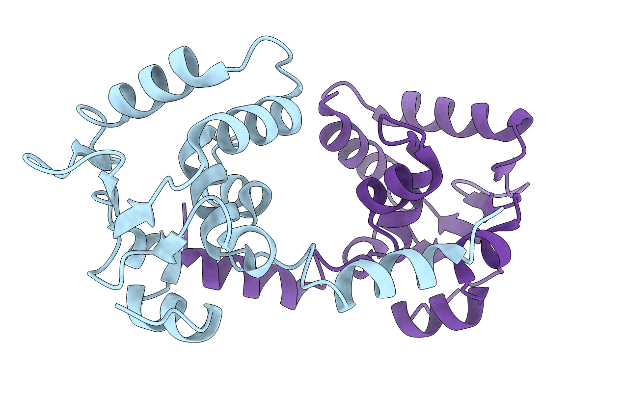
CRYSTAL STRUCTURE OF AN INHIBITED FRAGMENT OF Ets-1
Biological Source:
Source Organism:
PDB Version:
Deposition Date:
2002-08-06
Release Date:
2002-12-11
Method Details:
Experimental Method:
Resolution:
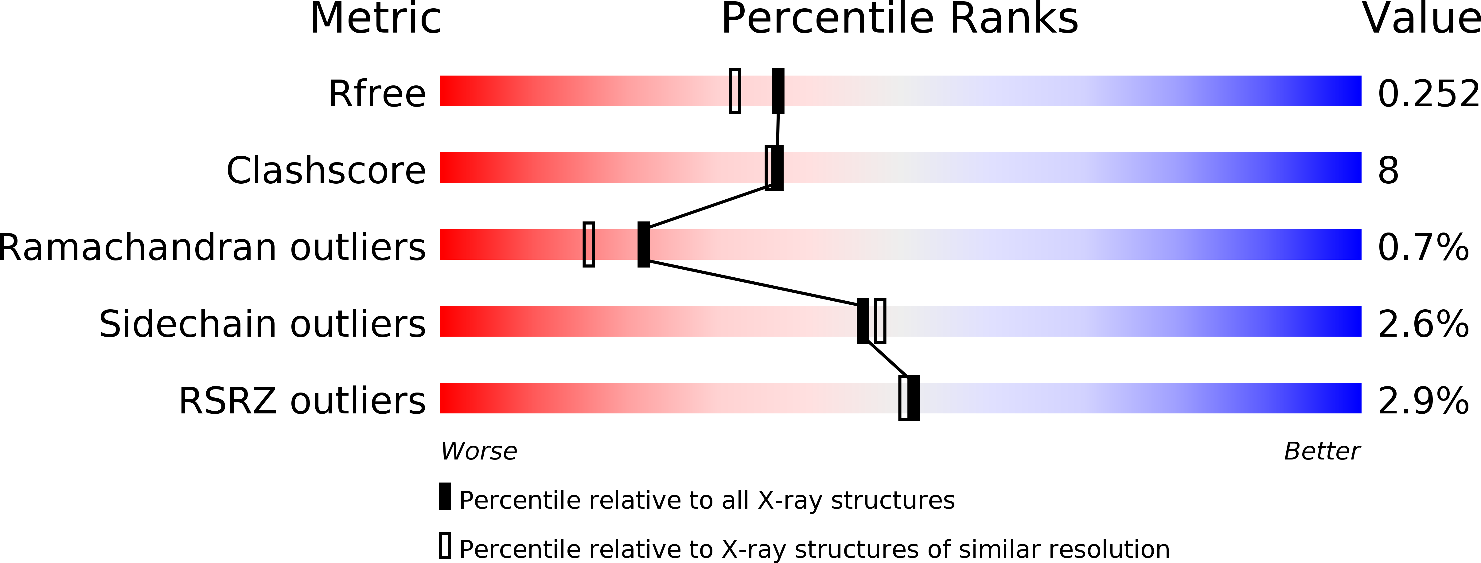
2.00 Å
R-Value Free:
0.23
R-Value Work:
0.20
Space Group:
P 21 21 21
Macromolecular Entities
Polymer Type:polypeptide(L)
Description:C-ets-1 protein
Chain IDs:A, B
Chain Length:141
Number of Molecules:2
Biological Source:Mus musculus
Primary Citation
Structural Analysis of the Autoinhibition of Ets-1 and Its Role in Protein Partnerships
J.Biol.Chem. 277 45529 45536 (2002)
PMID: 12221090 DOI: 10.1074/jbc.M206327200

Abstact

The DNA-binding activity of the eukaryotic transcription factor Ets-1 (E26 avian erythroblastosis virus oncogene-E twenty-six) is negatively regulated by inhibitory regions that flank the ETS domain. Based on the results of solution studies, these N- and C-terminal inhibitory regions have been proposed to pack against the ETS domain and form an autoinhibitory module whose N terminus partially unfolds upon binding of Ets-1 to DNA. Mutations that disrupt autoinhibition of DNA binding also cause a structural change in the inhibitory region. We report here a crystallographic study of fragments of Ets-1 that provide structural details of the inhibitory module and the structural transition that accompanies DNA binding. The structures of free and DNA-bound Ets-1 fragments containing the ETS domain and the inhibitory regions confirm that the N-terminal inhibitory region contains two alpha-helices one of which unfolds upon Ets-1 binding to DNA. The observations from the crystal structure, coupled with mutagenesis experiments, allow us to propose a model for the inhibited form of Ets-1 and lend insight into the flexible interaction between Ets-1 and the acute myeloid leukemia 1 protein, AML1 (RUNX1).

Legend

Protein

Chemical

Disease

Primary Citation of related structures