Deposition Date
2002-08-06
Release Date
2003-03-04
Last Version Date
2024-11-13
Entry Detail
PDB ID:
1MCX
Keywords:
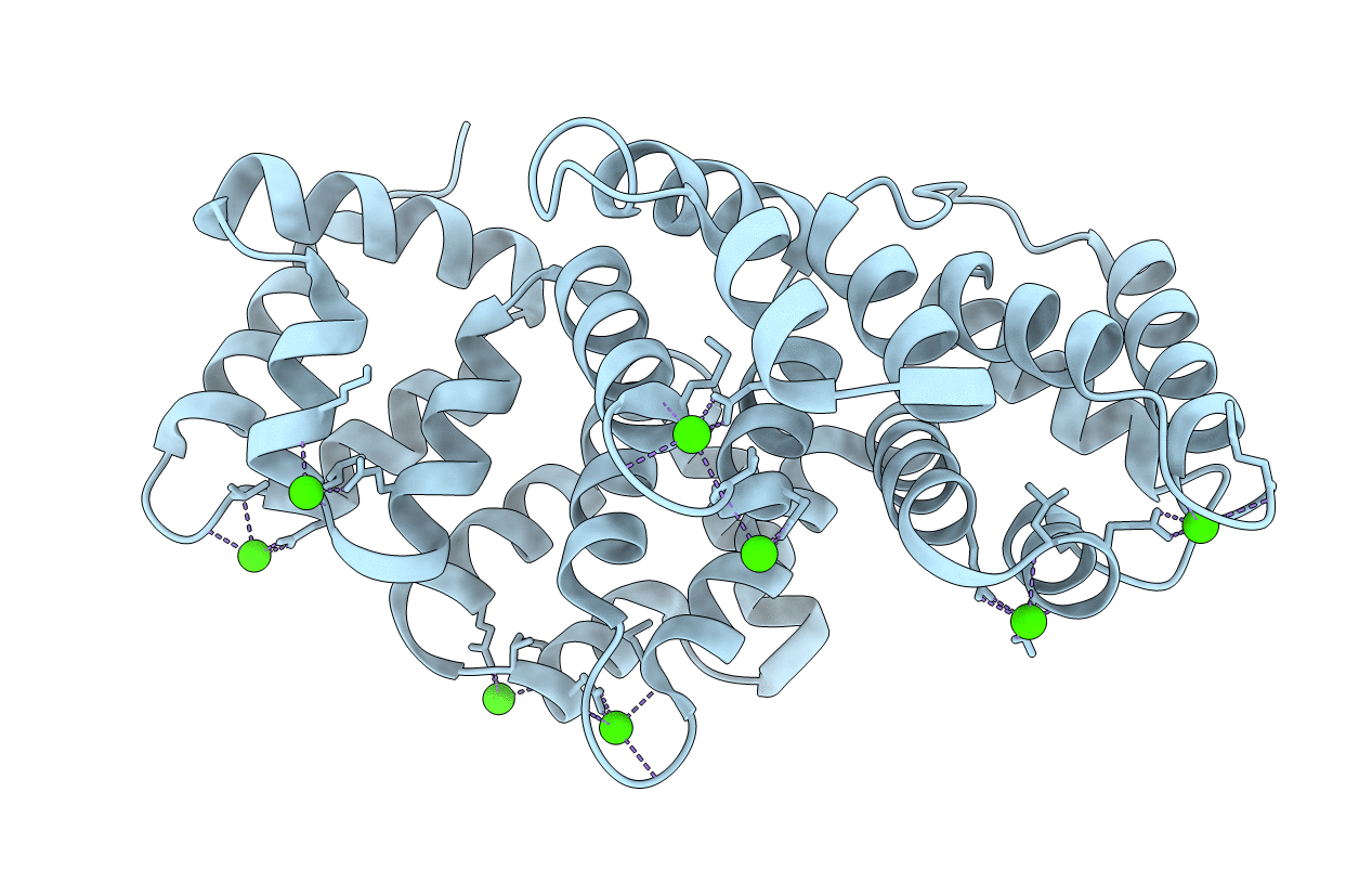
Title:
STRUCTURE OF FULL-LENGTH ANNEXIN A1 IN THE PRESENCE OF CALCIUM
Biological Source:
Source Organism(s):
Sus scrofa (Taxon ID: 9823)
Expression System(s):
Method Details:
Experimental Method:
Resolution:
2.03 Å
R-Value Free:
0.23
R-Value Work:
0.19
Space Group:
P 41 21 2