Deposition Date
2002-08-05
Release Date
2003-03-18
Last Version Date
2024-02-14
Entry Detail
PDB ID:
1MC4
Keywords:
Title:
Crystal Structure of Aspartate-Semialdehyde dehydrogenase from Vibrio Cholerae El Tor
Biological Source:
Source Organism(s):
Vibrio cholerae (Taxon ID: 666)
Expression System(s):
Method Details:
Experimental Method:
Resolution:
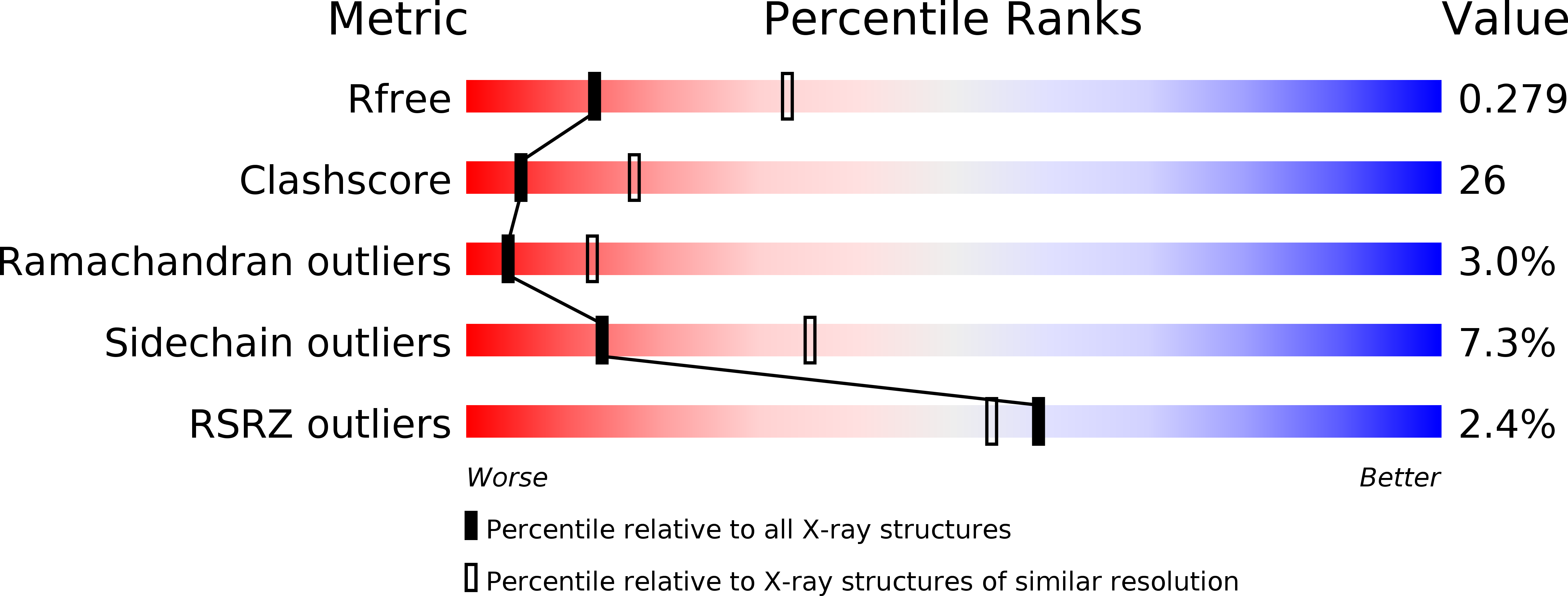
2.77 Å
R-Value Free:
0.28
R-Value Work:
0.21
Space Group:
P 43 3 2