Deposition Date
2002-08-02
Release Date
2003-06-10
Last Version Date
2024-02-14
Entry Detail
PDB ID:
1MB8
Keywords:
Title:
Crystal Structure of the actin binding domain of plectin
Biological Source:
Source Organism(s):
Homo sapiens (Taxon ID: 9606)
Expression System(s):
Method Details:
Experimental Method:
Resolution:
2.15 Å
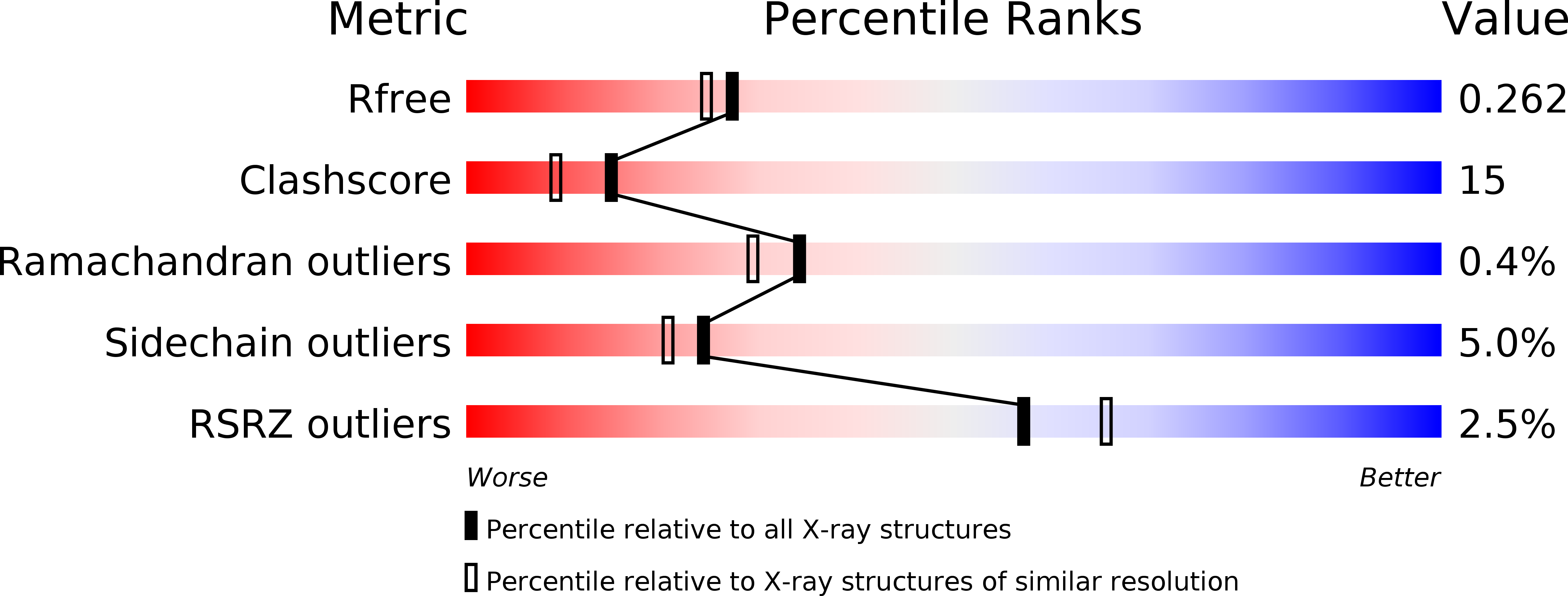
R-Value Free:
0.26
R-Value Work:
0.21
Space Group:
P 21 21 21