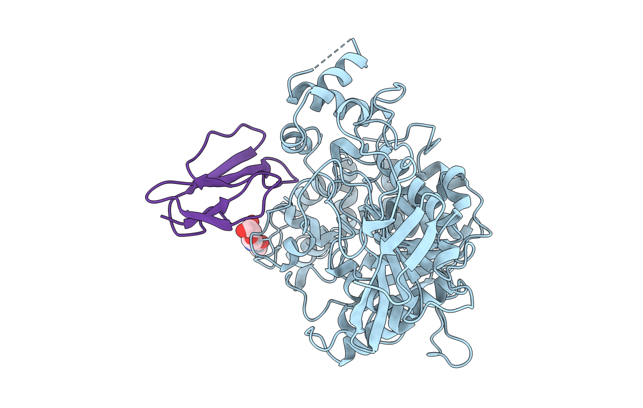
1MAH image
Deposition Date 1995-11-21
Release Date 1996-04-03
Last Version Date 2024-10-23
Entry Detail
PDB ID:
1MAH
Title:
FASCICULIN2-MOUSE ACETYLCHOLINESTERASE COMPLEX
Biological Source:
Source Organism:
Host Organism:
Method Details:
Experimental Method:
Resolution:
3.20 Å
R-Value Free:
0.29
R-Value Work:
0.18
R-Value Observed:
0.18
Space Group:
P 65 2 2
Macromolecular Entities
Structures with similar UniProt ID
Protein Blast
Polymer Type:polypeptide(L)
Molecule:ACETYLCHOLINESTERASE
Gene (Uniprot):Ache
Chain IDs:A
Chain Length:543
Number of Molecules:1
Biological Source:Mus musculus
Structures with similar UniProt ID
Protein Blast
Polymer Type:polypeptide(L)
Molecule:FASCICULIN 2
Chain IDs:B (auth: F)
Chain Length:61
Number of Molecules:1
Biological Source:Dendroaspis angusticeps
Modified Residue
Compound ID Chain ID Parent Comp ID Details 2D Image
ASN A ASN GLYCOSYLATION SITE
Ligand Molecules
Primary Citation
Acetylcholinesterase inhibition by fasciculin: crystal structure of the complex.
Cell(Cambridge,Mass.) 83 503 512 (1995)
PMID: 8521480 DOI: 10.1016/0092-8674(95)90128-0

Abstact

The crystal structure of the snake toxin fasciculin, bound to mouse acetylcholinesterase (mAChE), at 3.2 A resolution reveals a synergistic three-point anchorage consistent with the picomolar dissociation constant of the complex. Loop II of fasciculin contains a cluster of hydrophobic residues that interact with the peripheral anionic site of the enzyme and sterically occlude substrate access to the catalytic site. Loop I fits in a crevice near the lip of the gorge to maximize the surface area of contact of loop II at the gorge entry. The fasciculin core surrounds a protruding loop on the enzyme surface and stabilizes the whole assembly. Upon binding of fasciculin, subtle structural rearrangements of AChE occur that could explain the observed residual catalytic activity of the fasciculin-enzyme complex.

Legend

Protein

Chemical

Disease

Primary Citation of related structures
Feedback Form
Name
Email
Institute
Feedback