1MA9 image
Deposition Date 2002-08-02
Release Date 2003-02-04
Last Version Date 2025-03-26
Entry Detail
PDB ID:
1MA9
Title:
Crystal structure of the complex of human vitamin D binding protein and rabbit muscle actin
Biological Source:
Source Organism:
Method Details:
Experimental Method:
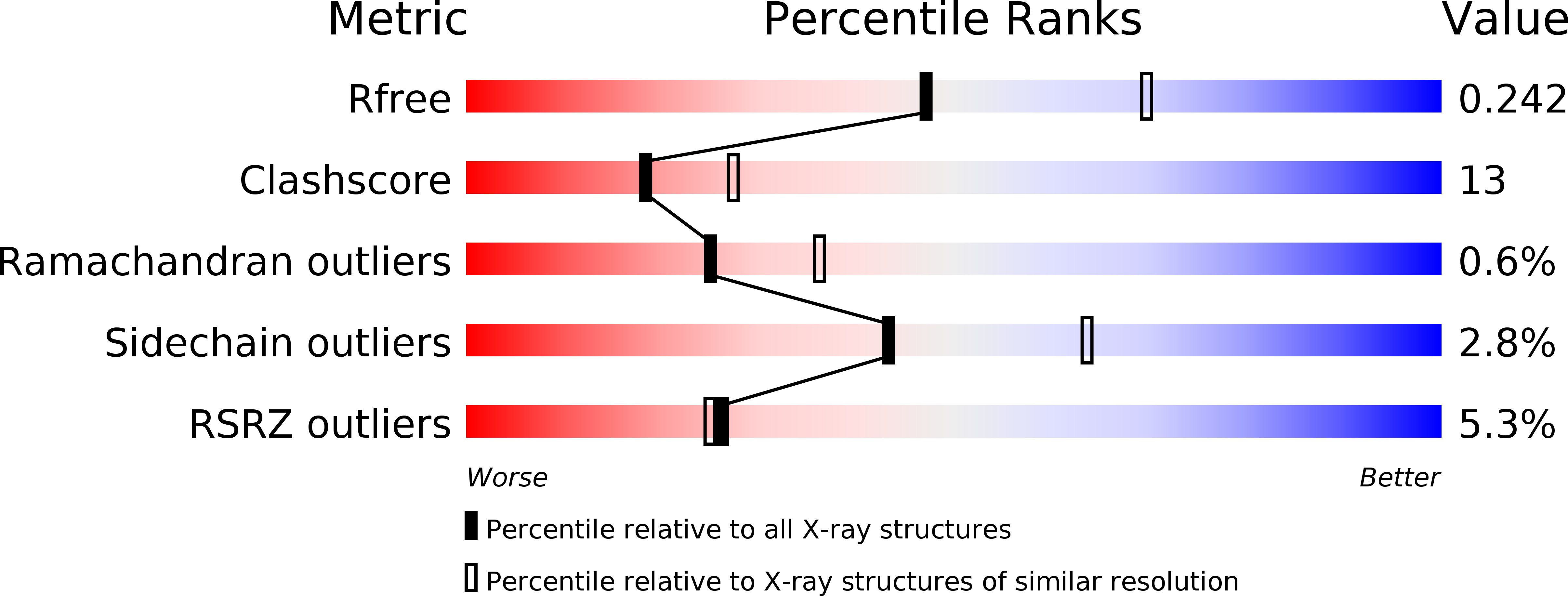
Resolution:
2.40 Å
R-Value Free:
0.25
R-Value Work:
0.2
R-Value Observed:
0.20
Space Group:
P 1 21 1
Macromolecular Entities
Polymer Type:polypeptide(L)
Molecule:Vitamin D-binding protein
Gene (Uniprot):GC
Chain IDs:A
Chain Length:458
Number of Molecules:1
Biological Source:Homo sapiens
Polymer Type:polypeptide(L)
Molecule:Actin, Alpha Skeletal Muscle
Gene (Uniprot):ACTA1
Chain IDs:B
Chain Length:375
Number of Molecules:1
Biological Source:Oryctolagus cuniculus
Modified Residue
Compound ID Chain ID Parent Comp ID Details 2D Image
HIC B HIS 4-METHYL-HISTIDINE
Primary Citation

Abstact

The multifunctional vitamin D binding protein (DBP) is an actin-sequestering protein present in blood. The crystal structure of the actin-DBP complex was determined at 2.4 A resolution. DBP binds to actin subdomains 1 and 3 and occludes the cleft at the interface between these subdomains. Most remarkably, DBP demonstrates an unusually large actin-binding interface, far exceeding the binding-interface areas reported for other actin-binding proteins such as profilin, DNase I and gelsolin. The fast-growing side of actin monomers is blocked completely through a perfect structural fit with DBP, demonstrating how DBP effectively interferes with actin-filament formation. It establishes DBP as the hitherto best actin-sequestering protein and highlights its key role in suppressing and preventing extracellular actin polymerization.

Legend

Protein

Chemical

Disease

Primary Citation of related structures