Deposition Date
2002-07-31
Release Date
2002-10-16
Last Version Date
2024-11-20
Entry Detail
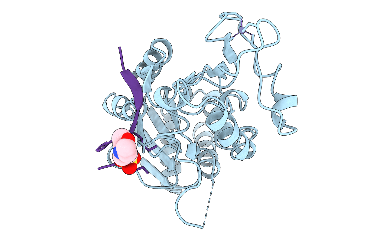
PDB ID:
1MA3
Keywords:
Title:
Structure of a Sir2 enzyme bound to an acetylated p53 peptide
Biological Source:
Source Organism(s):
Archaeoglobus fulgidus (Taxon ID: 2234)
Expression System(s):
Method Details:
Experimental Method:
Resolution:
2.00 Å
R-Value Free:
0.25
R-Value Work:
0.20
R-Value Observed:
0.21
Space Group:
P 21 21 21