1M7D image
Deposition Date 2002-07-19
Release Date 2003-07-22
Last Version Date 2024-10-16
Entry Detail
PDB ID:
1M7D
Keywords:
Title:
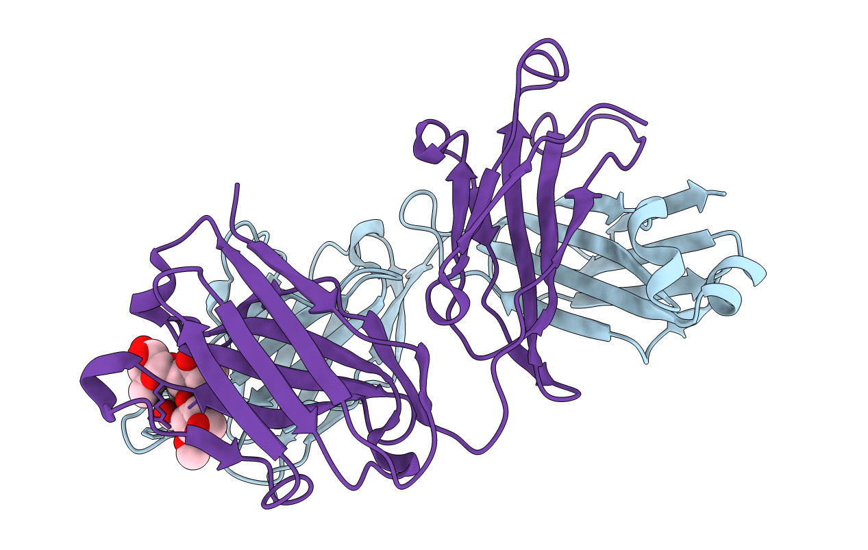
Crystal structure of a Monoclonal Fab Specific for Shigella flexneri Y Lipopolysaccharide complexed with a trisaccharide
Biological Source:
Source Organism:
Mus musculus (Taxon ID: 10090)
Host Organism:
Method Details:
Experimental Method:
Resolution:
2.30 Å
R-Value Free:
0.27
R-Value Work:
0.21
Space Group:
P 43 21 2
Macromolecular Entities
Polymer Type:polypeptide(L)
Molecule:light chain of the monoclonal antibody Fab SYA/J6
Chain IDs:A
Chain Length:215
Number of Molecules:1
Biological Source:Mus musculus
Polymer Type:polypeptide(L)
Molecule:heavy chain of the monoclonal antibody Fab SYA/J6
Chain IDs:B
Chain Length:220
Number of Molecules:1
Biological Source:Mus musculus
Primary Citation
Molecular Recognition of Oligosaccharide Epitopes by a Monoclonal Fab Specific for Shigella flexneri Y Lipopolysaccharide: X-ray Structures and Thermodynamics
Biochemistry 41 13575 13586 (2002)
PMID: 12427018 DOI: 10.1021/bi0261387

Abstact

The antigenic recognition of Shigella flexneri O-polysaccharide, which consists of a repeating unit ABCD [-->2)-alpha-L-Rhap-(1-->2)-alpha-L-Rhap-(1-->3)-alpha-L-Rhap-(1-->3)-beta-D-GlcpNAc-(1-->], by the monoclonal antibody SYA/J6 (IgG3, kappa) has been investigated by crystallographic analysis of the Fab domain and its two complexes with two antigen segments (a pentasaccharide Rha A-Rha B-Rha C-GlcNAc D-Rha A' and a modified trisaccharide Rha B-Rha C-GlcNAc D in which Rha C* is missing a C2-OH group). These complex structures, the first for a Fab specific for a periodic linear heteropolysaccharide, reveal a binding site groove (between the V(H) and V(L) domains) that makes polar and nonpolar contacts with all the sugar residues of the pentasaccharide. Both main-chain and side-chain atoms of the Fab are used in ligand binding. The charged side chain of Glu H50 of CDR H2 forms crucial hydrogen bonds to GlcNAc of the oligosaccharides. The modified trisaccharide is more buried and fits more snugly than the pentasaccharide. It also makes as many contacts (approximately 75) with the Fab as the pentasaccharide, including the same number of hydrogen bonds (eight, with four being identical). It is further engaged in more hydrophobic interactions than the pentasaccharide. These three features favorable to trisaccharide binding are consistent with the observation of a tighter complex with the trisaccharide than the pentasaccharide. Thermodynamic data demonstrate that the native tri- to pentasaccharides have free energies of binding in the range of 6.8-7.4 kcal mol(-1), and all but one of the hydrogen bonds to individual hydroxyl groups provide no more than approximately 0.7 kcal mol(-1). They further indicate that hydrophobic interactions make significant contributions to binding and, as the native epitope becomes larger across the tri-, tetra-, pentasaccharide series, entropy contributions to the free energy become dominant.

Legend

Protein

Chemical

Disease

Primary Citation of related structures