1M6T image
Deposition Date 2002-07-17
Release Date 2002-11-06
Last Version Date 2023-08-30
Entry Detail
PDB ID:
1M6T
Title:
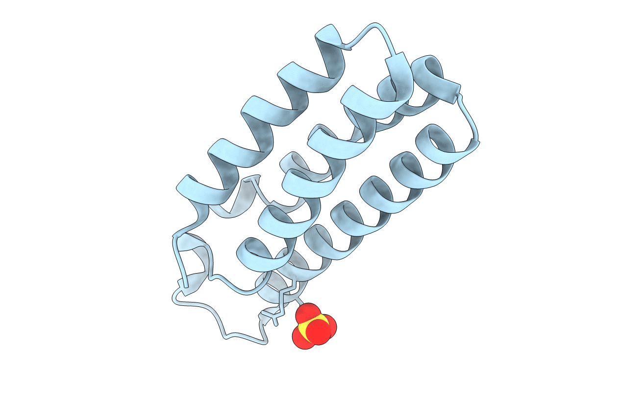
CRYSTAL STRUCTURE OF B562RIL, A REDESIGNED FOUR HELIX BUNDLE
Biological Source:
Source Organism(s):
Escherichia coli (Taxon ID: 562)
Expression System(s):
Method Details:
Experimental Method:
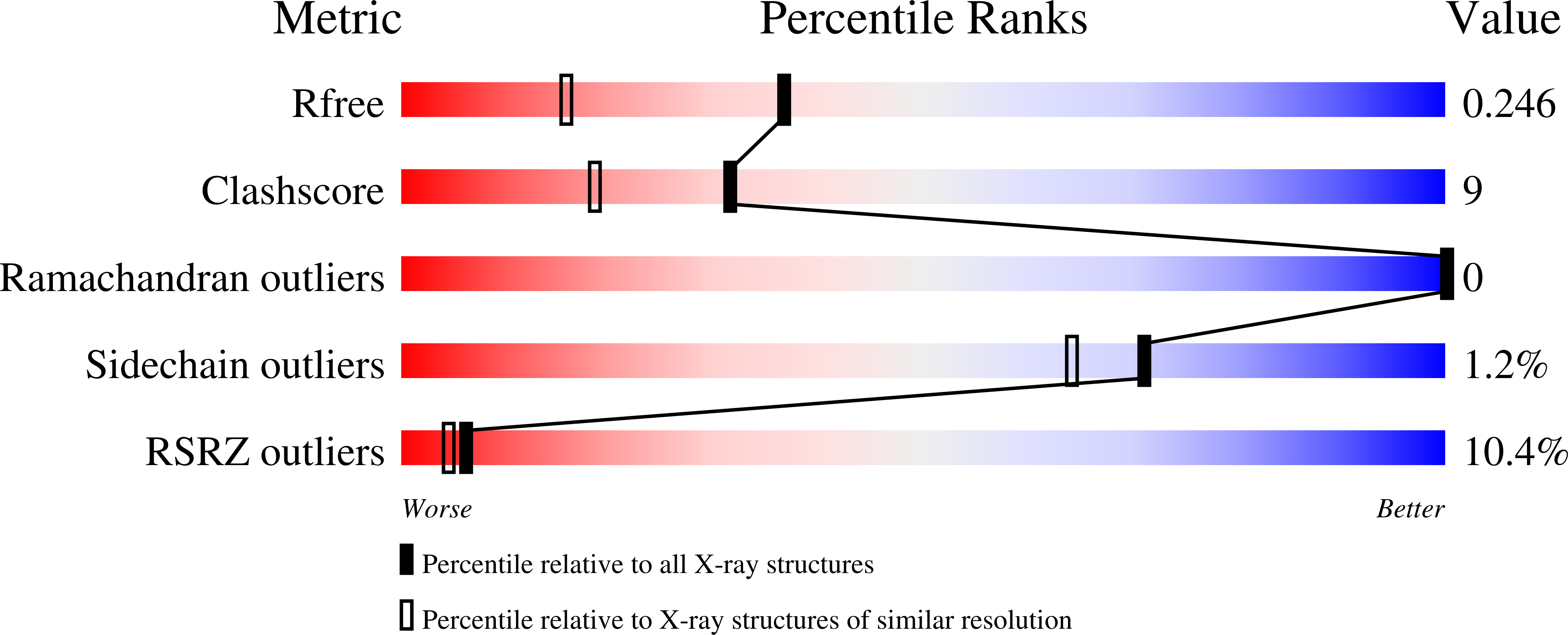
Resolution:
1.81 Å
R-Value Free:
0.24
R-Value Work:
0.21
Space Group:
C 2 2 21
Macromolecular Entities
Structures with similar UniProt ID
Protein Blast
Polymer Type:polypeptide(L)
Molecule:Soluble cytochrome b562
Gene (Uniprot):cybC
Mutagens:M7W,H102I,R106L
Chain IDs:A
Chain Length:106
Number of Molecules:1
Biological Source:Escherichia coli
Ligand Molecules
Primary Citation
Redesign of a Four-Helix Bundle Protein by Phage Display Coupled with Proteolysis and Structural Characterization by NMR and X-ray Crystallography
J.Mol.Biol. 323 253 262 (2002)
PMID: 12381319 DOI: 10.1016/S0022-2836(02)00884-7

Abstact

To test whether it is practical to use phage display coupled with proteolysis for protein design, we used this approach to convert a partially unfolded four-helix bundle protein, apocytochrome b(562), to a stably folded four-helix bundle protein. Four residues expected to form a hydrophobic core were mutated. One residue was changed to Trp to provide a fluorescence probe for studying the protein's physical properties and to partially fill the void left by the heme. The other three positions were randomly mutated. In addition, another residue in the region to be redesigned was substituted with Arg to provide a specific cutting site for protease Arg-c. This library of mutants was displayed on the surface of phage and challenged with protease Arg-c to select stably folded proteins. The consensus sequence that emerged from the selection included hydrophobic residues at only one of the three positions and non-hydrophobic residues at the other two. Nevertheless, the selected proteins were thermodynamically very stable. The structure of a selected protein was characterized using multi-dimensional NMR. All four helices were formed in the structure. Further, site-directed mutagenesis was used to change one of the two non-hydrophobic residues to a hydrophobic residue, which increased the stability of the protein, indicating that the selection result was not based solely on the protein's global stability and that local structural characteristics may also govern the selection. This conclusion is supported by the crystal structure of another mutant that has two hydrophobic residues substituted for the two non-hydrophobic residues. These results suggest that the hydrophobic interactions in the core are not sufficient to dictate the selection and that the location of the cutting site of the protease also influences the selection of structures.

Legend

Protein

Chemical

Disease

Primary Citation of related structures
Feedback Form
Name
Email
Institute
Feedback