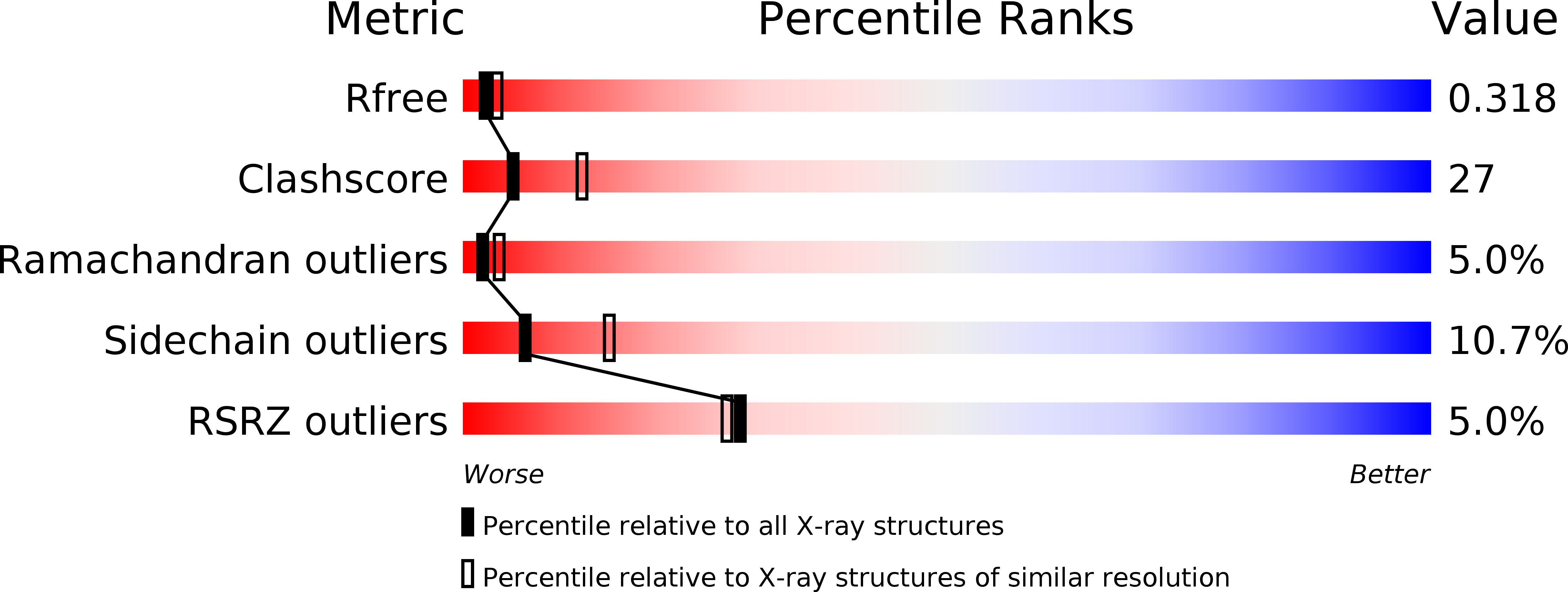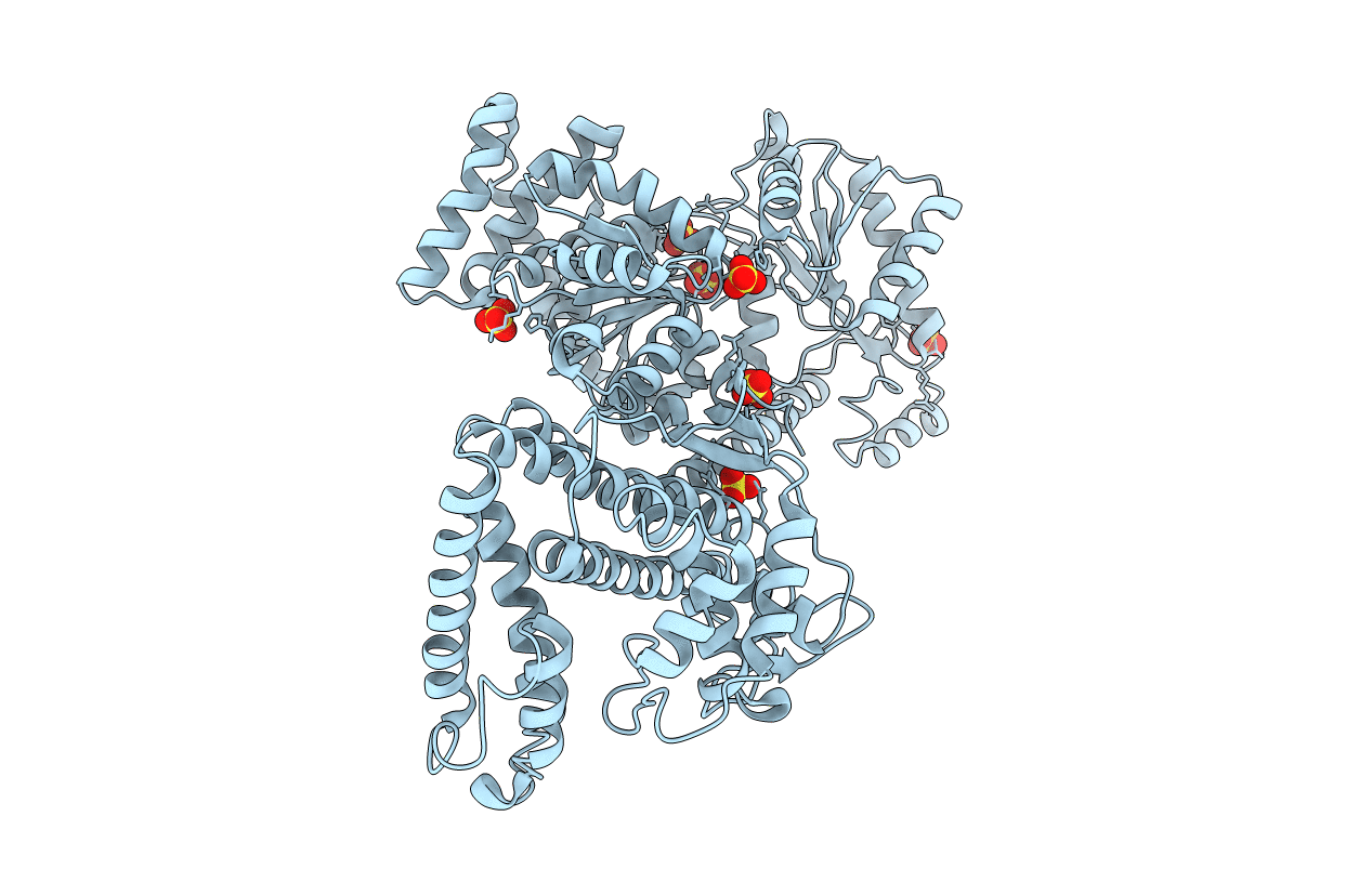
1M6N image
Deposition Date 2002-07-16
Release Date 2002-09-20
Last Version Date 2024-02-14
Entry Detail
PDB ID:
1M6N
Title:
Crystal structure of the SecA translocation ATPase from Bacillus subtilis
Biological Source:
Source Organism:
Method Details:
Experimental Method:
Resolution:
2.70 Å
R-Value Free:
0.30
R-Value Work:
0.22
R-Value Observed:
0.22
Space Group:
P 31 1 2
Macromolecular Entities
Polymer Type:polypeptide(L)
Molecule:Preprotein translocase secA
Gene (Uniprot):secA
Chain IDs:A
Chain Length:802
Number of Molecules:1
Biological Source:Bacillus subtilis
Ligand Molecules
Primary Citation
Nucleotide Control of Interdomain Interactions in the Conformational Reaction Cycle of SecA
Science 297 2018 2026 (2002)
PMID: 12242434 DOI: 10.1126/science.1074424

Abstact

The SecA adenosine triphosphatase (ATPase) mediates extrusion of the amino termini of secreted proteins from the eubacterial cytosol based on cycles of reversible binding to the SecYEG translocon. We have determined the crystal structure of SecA with and without magnesium-adenosine diphosphate bound to the high-affinity ATPase site at 3.0 and 2.7 angstrom resolution, respectively. Candidate sites for preprotein binding are located on a surface containing the SecA epitopes exposed to the periplasm upon binding to SecYEG and are thus positioned to deliver preprotein to SecYEG. Comparisons with structurally related ATPases, including superfamily I and II ATP-dependent helicases, suggest that the interaction geometry of the tandem motor domains in SecA is modulated by nucleotide binding, which is shown by fluorescence anisotropy experiments to reverse an endothermic domain-dissociation reaction hypothesized to gate binding to SecYEG.

Legend

Protein

Chemical

Disease

Primary Citation of related structures