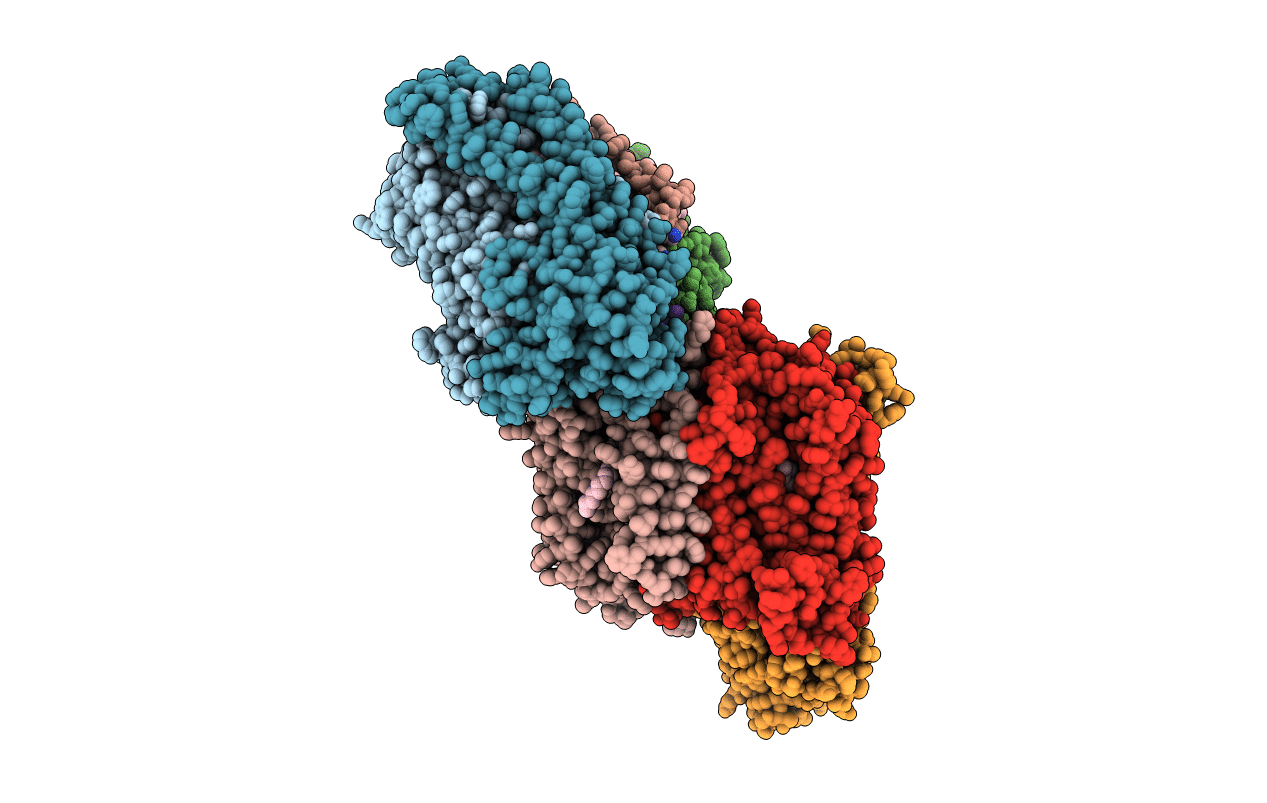
1M57 image
Entry Detail
PDB ID:
1M57
Keywords:
Title:
Structure of cytochrome c oxidase from Rhodobacter sphaeroides (EQ(I-286) mutant))
Biological Source:
Source Organism:
PDB Version:
Deposition Date:
2002-07-08
Release Date:
2002-08-28
Method Details:
Experimental Method:
Resolution:
3.00 Å
R-Value Free:
0.32
R-Value Work:
0.29
R-Value Observed:
0.29
Space Group:
H 3
Macromolecular Entities
Polymer Type:polypeptide(L)
Description:CYTOCHROME C OXIDASE
Mutations:E286Q
Chain IDs:A, E (auth: G)
Chain Length:566
Number of Molecules:2
Biological Source:Rhodobacter sphaeroides
Polymer Type:polypeptide(L)
Description:CYTOCHROME C OXIDASE
Chain IDs:B, F (auth: H)
Chain Length:264
Number of Molecules:2
Biological Source:Rhodobacter sphaeroides
Polymer Type:polypeptide(L)
Description:CYTOCHROME C OXIDASE
Chain IDs:C, G (auth: I)
Chain Length:266
Number of Molecules:2
Biological Source:Rhodobacter sphaeroides
Polymer Type:polypeptide(L)
Description:CYTOCHROME C OXIDASE
Chain IDs:D, H (auth: J)
Chain Length:51
Number of Molecules:2
Biological Source:Rhodobacter sphaeroides
Primary Citation
The X-ray crystal structures of wild-type and EQ(I-286) mutant cytochrome c oxidases from Rhodobacter sphaeroides.
J.Mol.Biol. 321 329 339 (2002)
PMID: 12144789 DOI: 10.1016/S0022-2836(02)00619-8

Abstact

The structure of cytochrome c oxidase from Rhodobacter sphaeroides has been solved at 2.3/2.8A (anisotropic resolution). This high-resolution structure revealed atomic details of a bacterial terminal oxidase including water molecule positions and a potential oxygen pathway, which has not been reported in other oxidase structures. A comparative study of the wild-type and the EQ(I-286) mutant enzyme revealed structural rearrangements around E(I-286) that could be crucial for proton transfer in this enzyme. In the structure of the mutant enzyme, EQ(I-286), which cannot transfer protons during oxygen reduction, the side-chain of Q(I-286) does not have the hydrogen bond to the carbonyl oxygen of M(I-107) that is seen in the wild-type structure. Furthermore, the Q(I-286) mutant has a different arrangement of water molecules and residues in the vicinity of the Q side-chain. These differences between the structures could reflect conformational changes that take place upon deprotonation of E(I-286) during turnover of the wild-type enzyme, which could be part of the proton-pumping machinery of the enzyme.

Legend

Protein

Chemical

Disease

Primary Citation of related structures