1M4J image
Entry Detail
PDB ID:
1M4J
Title:
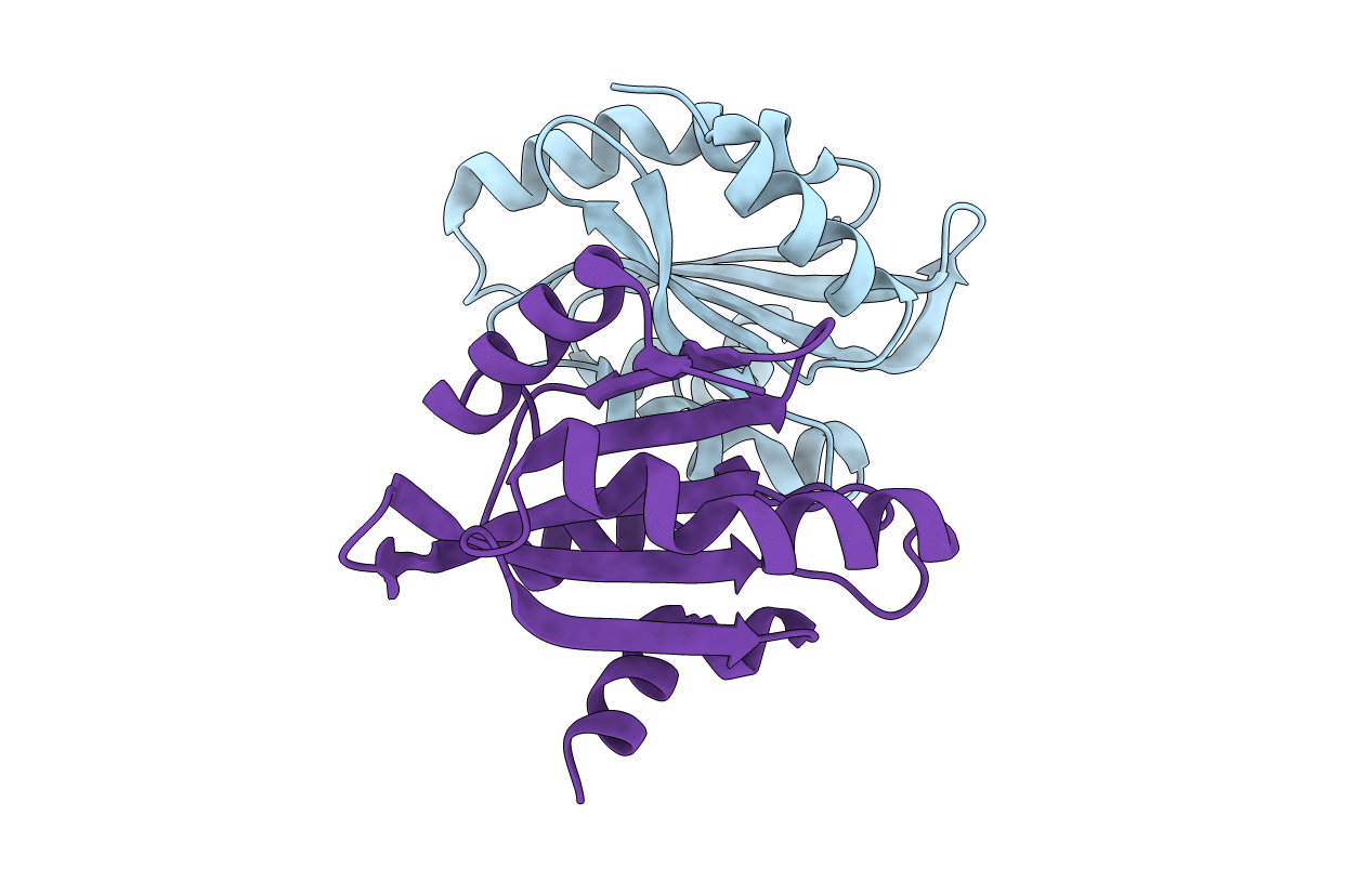
CRYSTAL STRUCTURE OF THE N-TERMINAL ADF-H DOMAIN OF MOUSE TWINFILIN ISOFORM-1
Biological Source:
Source Organism:
PDB Version:
Deposition Date:
2002-07-03
Release Date:
2002-11-13
Method Details:
Experimental Method:
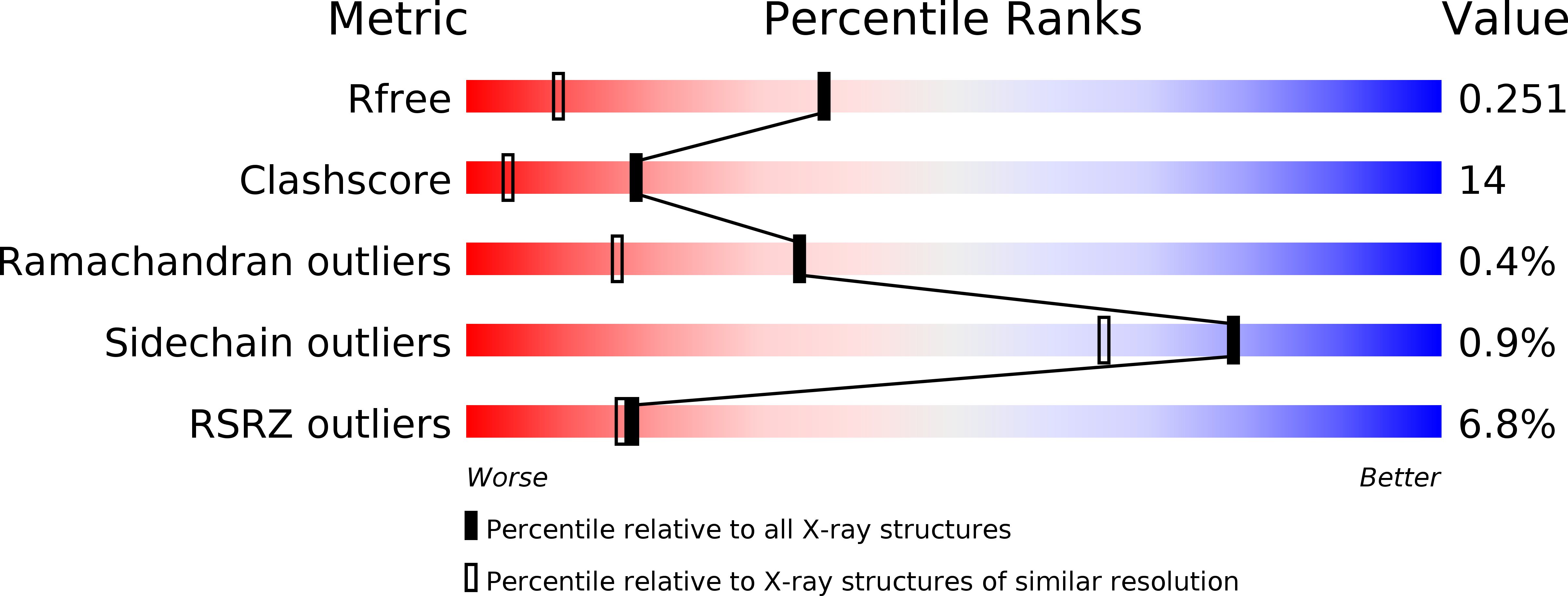
Resolution:
1.60 Å
R-Value Free:
0.25
R-Value Work:
0.23
R-Value Observed:
0.23
Space Group:
P 21 21 21
Macromolecular Entities
Polymer Type:polypeptide(L)
Description:A6 gene product
Chain IDs:A, B
Chain Length:142
Number of Molecules:2
Biological Source:Mus musculus
Primary Citation
Structural Conservation Between the Actin Monomer-binding Sites of Twinfilin and Actin-depolymerizing Factor (ADF)/Cofilin
J.Biol.Chem. 277 43089 43095 (2002)
PMID: 12207032 DOI: 10.1074/jbc.M208225200

Abstact

Twinfilin is an evolutionarily conserved actin monomer-binding protein that regulates cytoskeletal dynamics in organisms from yeast to mammals. It is composed of two actin-depolymerization factor homology (ADF-H) domains that show approximately 20% sequence identity to ADF/cofilin proteins. In contrast to ADF/cofilins, which bind both G-actin and F-actin and promote filament depolymerization, twinfilin interacts only with G-actin. To elucidate the molecular mechanisms of twinfilin-actin monomer interaction, we determined the crystal structure of the N-terminal ADF-H domain of twinfilin and mapped its actin-binding site by site-directed mutagenesis. This domain has similar overall structure to ADF/cofilins, and the regions important for actin monomer binding in ADF/cofilins are especially well conserved in twinfilin. Mutagenesis studies show that the N-terminal ADF-H domain of twinfilin and ADF/cofilins also interact with actin monomers through similar interfaces, although the binding surface is slightly extended in twinfilin. In contrast, the regions important for actin-filament interactions in ADF/cofilins are structurally different in twinfilin. This explains the differences in actin-interactions (monomer versus filament binding) between twinfilin and ADF/cofilins. Taken together, our data show that the ADF-H domain is a structurally conserved actin-binding motif and that relatively small structural differences at the actin interfaces of this domain are responsible for the functional variation between the different classes of ADF-H domain proteins.

Legend

Protein

Chemical

Disease

Primary Citation of related structures