1M4H image
Deposition Date 2002-07-02
Release Date 2002-08-28
Last Version Date 2024-11-20
Entry Detail
PDB ID:
1M4H
Title:
Crystal Structure of Beta-secretase complexed with Inhibitor OM00-3
Biological Source:
Source Organism:
Host Organism:
Method Details:
Experimental Method:
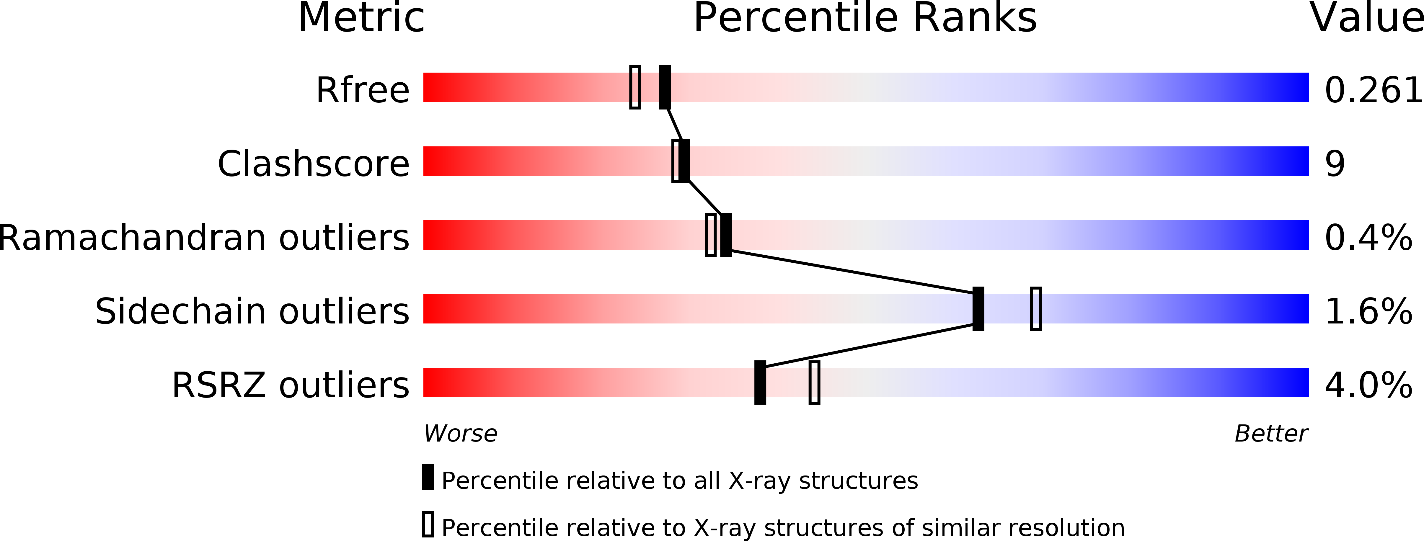
Resolution:
2.10 Å
R-Value Free:
0.27
R-Value Work:
0.21
Space Group:
P 21 21 21
Macromolecular Entities
Polymer Type:polypeptide(L)
Molecule:beta-Secretase
Gene (Uniprot):BACE1
Chain IDs:A, B
Chain Length:391
Number of Molecules:2
Biological Source:Homo sapiens
Polymer Type:polypeptide(L)
Molecule:Inhibitor OM00-3
Chain IDs:C, D
Chain Length:7
Number of Molecules:2
Biological Source:synthetic construct
Peptide-like Molecules
PRD_000289
Primary Citation
Crystal Structure of Memapsin 2 (beta-Secretase) in Complex with Inhibitor OM00-3
Biochemistry 41 10963 10967 (2002)
PMID: 12206667 DOI: 10.1021/bi026232n

Abstact

The structure of the catalytic domain of human memapsin 2 bound to an inhibitor OM00-3 (Glu-Leu-Asp-LeuAla-Val-Glu-Phe, K(i) = 0.3 nM, the asterisk denotes the hydroxyethylene transition-state isostere) has been determined at 2.1 A resolution. Uniquely defined in the structure are the locations of S(3)' and S(4)' subsites, which were not identified in the previous structure of memapsin 2 in complex with the inhibitor OM99-2 (Glu-Val-Asn-LeuAla-Ala-Glu-Phe, K(i) = 1 nM). Different binding modes for the P(2) and P(4) side chains are also observed. These new structural elements are useful for the design of new inhibitors. The structural and kinetic data indicate that the replacement of the P(2)' alanine in OM99-2 with a valine in OM00-3 stabilizes the binding of P(3)' and P(4)'.

Legend

Protein

Chemical

Disease

Primary Citation of related structures