Deposition Date
2002-06-28
Release Date
2003-01-21
Last Version Date
2024-02-14
Entry Detail
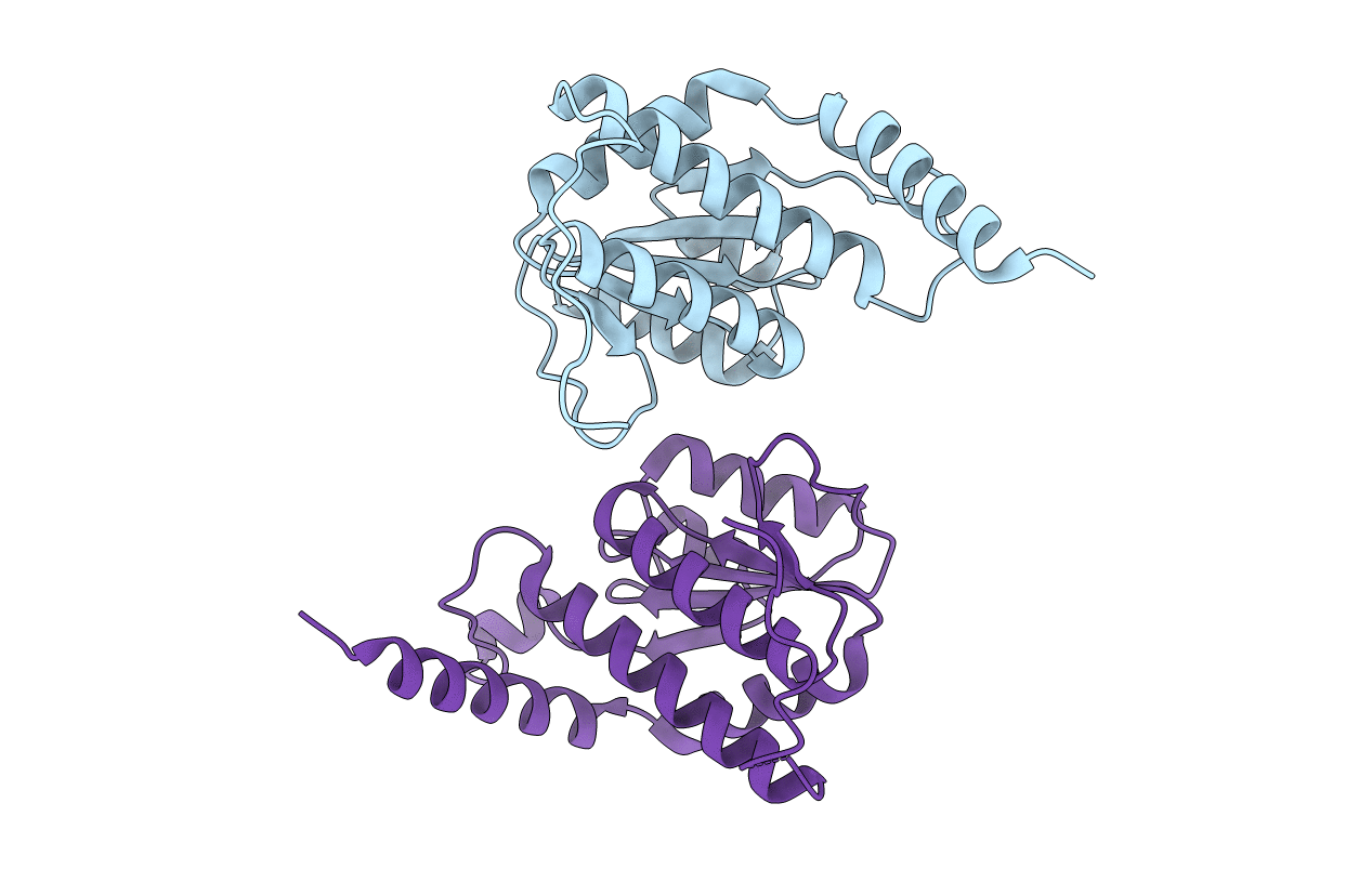
PDB ID:
1M3S
Keywords:
Title:
Crystal structure of YckF from Bacillus subtilis
Biological Source:
Source Organism(s):
Bacillus subtilis (Taxon ID: 1423)
Expression System(s):
Method Details:
Experimental Method:
Resolution:
1.95 Å
R-Value Free:
0.22
R-Value Work:
0.18
R-Value Observed:
0.18
Space Group:
P 65 2 2