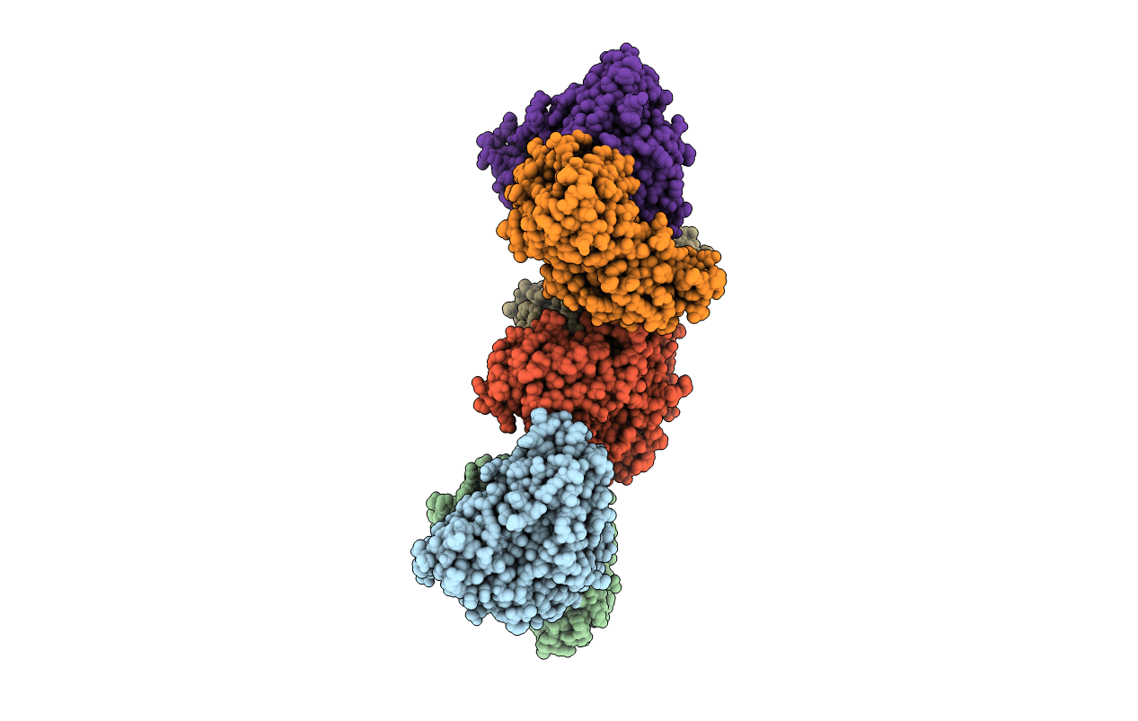
1M32 image
Deposition Date 2002-06-26
Release Date 2002-11-20
Last Version Date 2025-03-26
Entry Detail
PDB ID:
1M32
Keywords:
Title:
Crystal Structure of 2-aminoethylphosphonate Transaminase
Biological Source:
Source Organism:
Host Organism:
Method Details:
Experimental Method:
Resolution:
2.20 Å
R-Value Free:
0.2
R-Value Work:
0.16
R-Value Observed:
0.17
Space Group:
P 1 21 1
Macromolecular Entities
Polymer Type:polypeptide(L)
Molecule:2-aminoethylphosphonate-pyruvate aminotransferase
Gene (Uniprot):phnW
Chain IDs:A, B, C, D, E, F
Chain Length:366
Number of Molecules:6
Biological Source:Salmonella typhimurium
Modified Residue
Compound ID Chain ID Parent Comp ID Details 2D Image
MSE A MET SELENOMETHIONINE
Primary Citation
Degradation Pathway of the Phosphonate Ciliatine: Crystal Structure of 2-Aminoethylphosphonate Transaminase
Biochemistry 41 13162 13169 (2002)
PMID: 12403617 DOI: 10.1021/bi026231v

Abstact

Phosphonates allow certain organisms to thrive in otherwise hostile environments, and 2-aminoethylphosphonate (AEP) is a precursor of many cellular phosphonates. AEP transaminase (AEPT) is an enzyme essential to phosphonate synthesis and degradation pathways. The crystal structure of AEP transaminase was determined by multiwavelength anomalous diffraction of 66 selenium atoms. The refined structure at 2.2 A resolution revealed an overall fold and active site location similar to those of the dimeric, two-domain structure of type I aminotransferases. The active site contains a cofactor, pyridoxal 5'-phosphate (PLP), and the product phosphonoacetaldehyde. Comparison with other type I aminotransferase structures shows that the PLP-protein interactions are conserved. Modeling of bound substrates and products reveals the structural basis for AEP recognition and the stereospecificity of proton elimination at the alpha-carbon and indicates conformational changes along the reaction pathway.

Legend

Protein

Chemical

Disease

Primary Citation of related structures