Deposition Date
2002-06-20
Release Date
2002-09-11
Last Version Date
2023-10-25
Entry Detail
PDB ID:
1M20
Keywords:
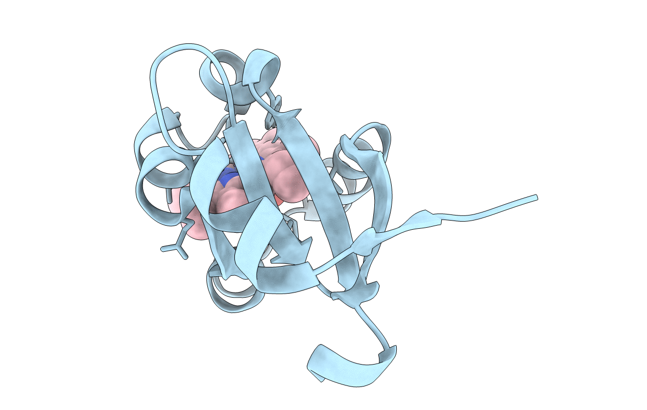
Title:
Crystal Structure of F35Y Mutant of Trypsin-solubilized Fragment of Cytochrome b5
Biological Source:
Source Organism(s):
Bos taurus (Taxon ID: 9913)
Expression System(s):
Method Details:
Experimental Method:
Resolution:
1.80 Å
R-Value Free:
0.22
R-Value Work:
0.19
R-Value Observed:
0.19
Space Group:
C 1 2 1