1M0V image
Entry Detail
PDB ID:
1M0V
Keywords:
Title:
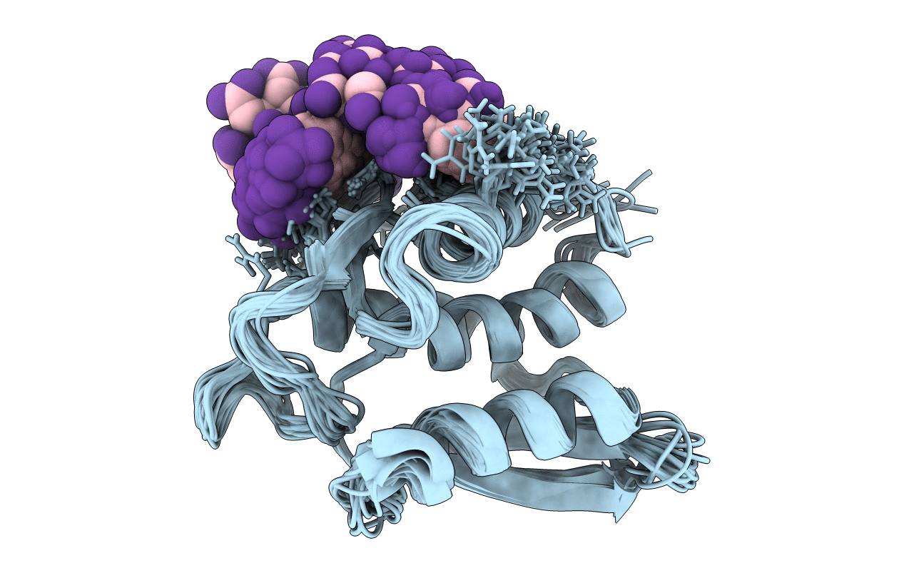
NMR STRUCTURE OF THE TYPE III SECRETORY DOMAIN OF YERSINIA YOPH COMPLEXED WITH THE SKAP-HOM PHOSPHO-PEPTIDE N-acetyl-DEpYDDPF-NH2
Biological Source:
Source Organism:
Host Organism:
PDB Version:
Deposition Date:
2002-06-14
Release Date:
2002-07-24
Method Details:
Experimental Method:
Conformers Calculated:
360
Conformers Submitted:
20
Selection Criteria:
structures with the least restraint violations
Macromolecular Entities
Polymer Type:polypeptide(L)
Description:PROTEIN-TYROSINE PHOSPHATASE YOPH
Chain IDs:A
Chain Length:136
Number of Molecules:1
Biological Source:Yersinia pseudotuberculosis
Polymer Type:polypeptide(L)
Description:SKAP55 homologue
Chain IDs:B
Chain Length:9
Number of Molecules:1
Biological Source:
Modified Residue
Compound ID Chain ID Parent Comp ID Details 2D Image
PTR B TYR O-PHOSPHOTYROSINE
Ligand Molecules
Primary Citation
Solution structure and phosphopeptide binding to the N-terminal domain of Yersinia YopH: comparison with a crystal structure
Biochemistry 41 11425 11437 (2002)
PMID: 12234185 DOI: 10.1021/bi026333l

Abstact

Virulence of pathogenic bacteria of the genus Yersinia requires the injection of six effector proteins into the cytoplasm of host cells. The amino-terminal domain of one of these effectors, the tyrosine phosphatase YopH, is essential for translocation of YopH, as well as for targeting it to phosphotyrosine-containing substrates of the type pYxxP. We report the high-resolution solution structure of the N-terminal domain (residues 1-129) from the Yersinia pseudotuberculosis YopH (YopH-NT) in complex with N-acetyl-DEpYDDPF-NH(2), a peptide derived from an in vivo protein substrate. In contrast to the domain-swapped dimer observed in a crystal structure of the same protein (Smith, C. L., Khandelwal, P., Keliikuli, K., Zuiderweg, E. R. P., and Saper, M. A. (2001) Mol. Microbiol. 42, 967-979), YopH-NT is monomeric in solution. The peptide binding site is located on a beta-hairpin that becomes the crossover point in the dimer structure. The binding site has several characteristics that are reminiscent of SH2 domains, which also bind to pYxxP sequences.

Legend

Protein

Chemical

Disease

Primary Citation of related structures