1M0D image
Deposition Date 2002-06-12
Release Date 2002-07-10
Last Version Date 2024-02-14
Entry Detail
PDB ID:
1M0D
Keywords:
Title:
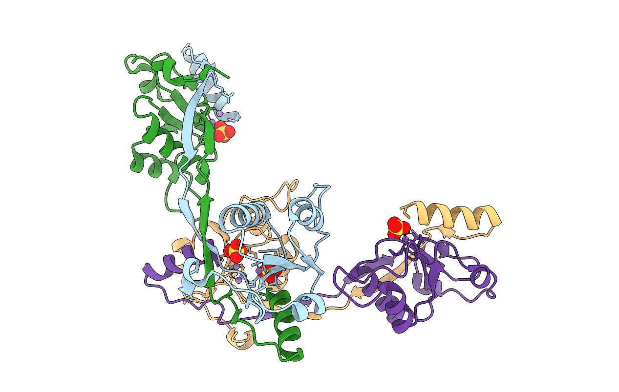
Crystal Structure of Bacteriophage T7 Endonuclease I with a Wild-Type Active Site and Bound Manganese Ions
Biological Source:
Source Organism:
Method Details:
Experimental Method:
Resolution:
1.90 Å
R-Value Free:
0.23
R-Value Work:
0.21
R-Value Observed:
0.21
Space Group:
P 21 21 2
Macromolecular Entities
Polymer Type:polypeptide(L)
Molecule:Endodeoxyribonuclease I
Gene (Uniprot):3
Chain IDs:A, B, C, D
Chain Length:138
Number of Molecules:4
Biological Source:Enterobacteria phage T7
Primary Citation
Metal ions bound at the active site of the junction-resolving enzyme T7 endonuclease I.
EMBO J. 21 3505 3515 (2002)
PMID: 12093751 DOI: 10.1093/emboj/cdf337

Abstact

T7 endonuclease I is a nuclease that is selective for the structure of the four-way DNA junction. The active site is similar to those of a number of restriction enzymes. We have solved the crystal structure of endonuclease I with a wild-type active site. Diffusion of manganese ions into the crystal revealed two peaks of electron density per active site, defining two metal ion-binding sites. Site 1 is fully occupied, and the manganese ion is coordinated by the carboxylate groups of Asp55 and Glu65, and the main chain carbonyl of Thr66. Site 2 is partially occupied, and the metal ion has a single protein ligand, the remaining carboxylate oxygen atom of Asp55. Isothermal titration calorimetry showed the sequential exothermic binding of two manganese ions in solution, with dissociation constants of 0.58 +/- 0.019 and 14 +/- 1.5 mM. These results are consistent with a two metal ion mechanism for the cleavage reaction, in which the hydrolytic water molecule is contained in the first coordination sphere of the site 1-bound metal ion.

Legend

Protein

Chemical

Disease

Primary Citation of related structures