1LZY image
Deposition Date 1995-01-09
Release Date 1995-02-27
Last Version Date 2024-10-23
Entry Detail
PDB ID:
1LZY
Title:
X-RAY STRUCTURE OF TURKEY EGG LYSOZYME COMPLEX WITH DI-N-ACETYLCHITOBIOSE. RECOGNITION AND BINDING OF ALPHA-ANOMERIC FORM
Biological Source:
Source Organism:
Method Details:
Experimental Method:
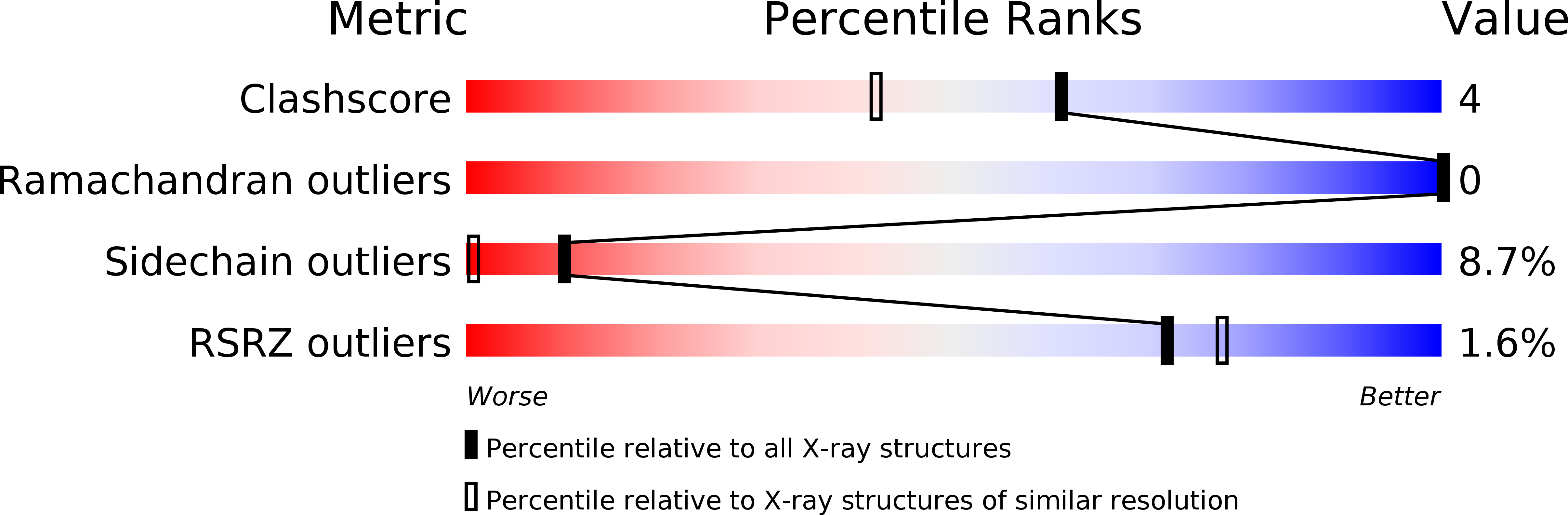
Resolution:
1.55 Å
R-Value Work:
0.17
R-Value Observed:
0.17
Space Group:
P 1 21 1
Macromolecular Entities
Polymer Type:polypeptide(L)
Molecule:TURKEY EGG WHITE LYSOZYME
Gene (Uniprot):LYZ
Chain IDs:A
Chain Length:129
Number of Molecules:1
Biological Source:Meleagris gallopavo
Primary Citation
X-ray structure of monoclinic turkey egg lysozyme at 1.3 A resolution.
Acta Crystallogr.,Sect.D 49 497 504 (1993)
PMID: 15299509 DOI: 10.1107/S0907444993005542

Abstact

Monoclinic crystals of turkey egg lysozyme (TEL, E.C. 3.2.1.17) were obtained from 2.2 M ammonium sulfate solution at pH 4.2. They belong to space group P2(1) with unit-cell dimensions a = 38.07, b = 33.20, c = 46.12 A and beta = 110.1 degrees, and contain one molecule in the asymmetric unit (V(m) = 1.91 A(3) Da(-1)). The three-dimensional structure of TEL was solved by the method of multiple isomorphous replacement with anomalous scattering. Area detector data to 1.5 A resolution from native and heavy-atom derivatives were used for the structure determination. The structure was refined by the simulated-annealing method with diffraction data of 10-1.30 A resolution. The conventional R factor was 0.189. The root-mean-square deviations from ideal bond distances and angles were 0.016 A and 2.9 degrees, respectively. The backbone structure of TEL is very similar to that of hen egg lysozyme (HEL) and the difference in seven amino-acid residues does not affect the basic folding of the polypeptide chain. Except for the region from Gly101 to Gly104, the geometry of the active-site cleft is conserved between TEL and HEL. The Gly101 residue is located at the end of the sugar-binding site and the structural change in this region between TEL and HEL is considered to be responsible for the difference in their enzymatic properties.

Legend

Protein

Chemical

Disease

Primary Citation of related structures