
Deposition Date
1994-09-14
Release Date
1995-04-20
Last Version Date
2024-10-09
Entry Detail
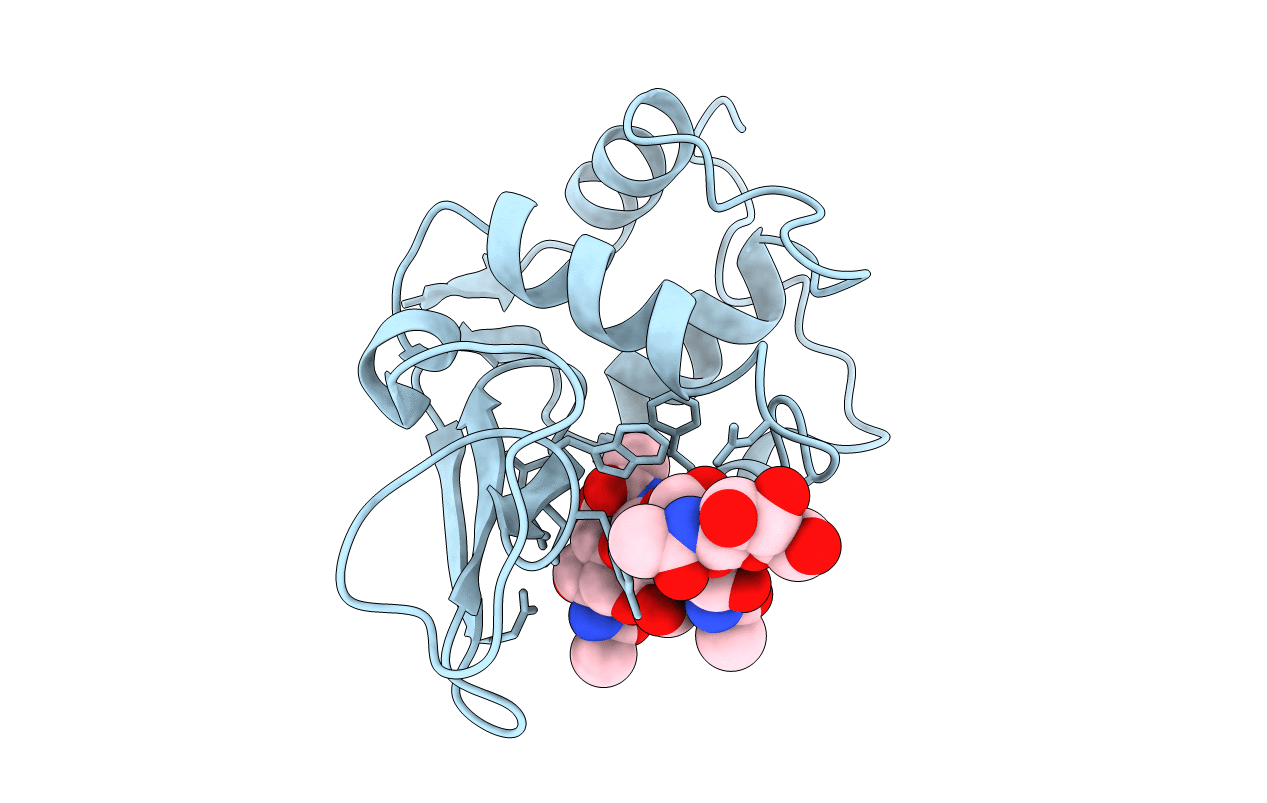
PDB ID:
1LZR
Keywords:
Title:
STRUCTURAL CHANGES OF THE ACTIVE SITE CLEFT AND DIFFERENT SACCHARIDE BINDING MODES IN HUMAN LYSOZYME CO-CRYSTALLIZED WITH HEXA-N-ACETYL-CHITOHEXAOSE AT PH 4.0
Biological Source:
Source Organism(s):
Homo sapiens (Taxon ID: 9606)
Method Details:
Experimental Method:
Resolution:
1.50 Å
R-Value Observed:
0.14
Space Group:
P 21 21 21
