1LYS image
Deposition Date 1993-12-03
Release Date 1994-04-30
Last Version Date 2024-11-20
Entry Detail
PDB ID:
1LYS
Title:
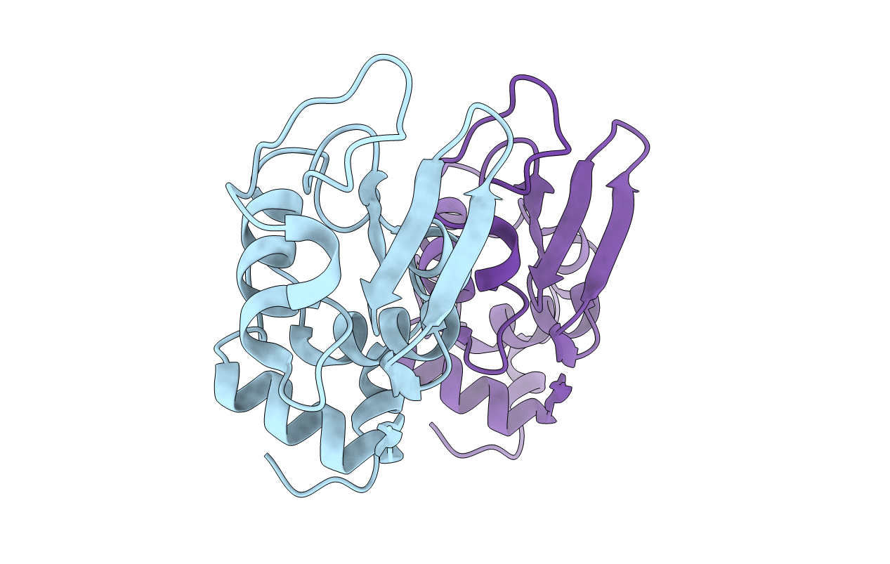
X-RAY STRUCTURE OF A MONOCLINIC FORM OF HEN EGG-WHITE LYSOZYME CRYSTALLIZED AT 313K. COMPARISON OF TWO INDEPENDENT MOLECULES
Biological Source:
Source Organism:
Gallus gallus (Taxon ID: 9031)
Method Details:
Experimental Method:
Resolution:
1.72 Å
R-Value Work:
0.18
R-Value Observed:
0.18
Space Group:
P 1 21 1
Macromolecular Entities
Structures with similar UniProt ID
Protein Blast
Polymer Type:polypeptide(L)
Molecule:HEN EGG WHITE LYSOZYME
Gene (Uniprot):LYZ
Chain IDs:A, B
Chain Length:129
Number of Molecules:2
Biological Source:Gallus gallus
Primary Citation
X-ray structure of a monoclinic form of hen egg-white lysozyme crystallized at 313 K. Comparison of two independent molecules.
Acta Crystallogr.,Sect.D 50 250 257 (1994)
PMID: 15299435 DOI: 10.1107/S0907444993013290

Abstact

A monoclinic crystal of hen egg lysozyme (HEL, E.C. 3.2.1.17) was obtained at 313 K from a 10%(w/v) NaCl solution at pH 7.6 containing 5%(v/v) 1-propanol. Cell dimensions were a = 27.23, b = 63.66, c = 59.12 A and beta = 92.9 degrees, and the space group was P2(1). The unit cell contains four molecules (V(m) = 1.79 A(3) Da(-1)). The structure was solved by the isomorphous replacement method with anomalous scattering followed by phase improvement by the solvent-flattening method. The refinement of the structure was carried out by the simulated-annealing method. The conventional R value was 0.187 for 18 260 reflections [|F(o)| > 3sigma(F)] in the resolution range 10-1.72 A. The r.m.s. deviations from the ideal bond distances and angles were 0.015 A and 3.0 degrees, respectively. The two molecules in the asymmetric unit are related by a translation of half a lattice unit along the a and c axes. The r.m.s. difference of equivalent C(alpha) atoms between the two molecules was 0.64 A and the largest difference was 3.57 A for Gly71. A significant structural change was observed in the regions of residues 45-50, 65-73 and 100-104. The residues 45-50, which connect two beta-strands, are shifted parallel to the beta-sheet plane between the two molecules. The residues 100-104 belong to the substrate-binding site (subsite A) and the high flexibility of this region may be responsible for the binding of the substrate and the release of reaction products.

Legend

Protein

Chemical

Disease

Primary Citation of related structures